Sara N. Bramlett
Ph.D.

Science Communicator  ·  Scientific Illustrator  ·  Neuropharmacologist  ·  Artist

Science is only as powerful as our ability to share it. As a neuropharmacologist turned visual communicator, I specialize in translating high-level concepts into engaging narratives. From digital molecular pathways to physical installations using resin, electronics, and mixed media, I create eye-catching visuals and immersive experiences that spark curiosity and simplify the complex. My personal motto is: create, by any means necessary

SciOpsDesign logo

Science, Art,
and Everything Between

I'm a neuropharmacologist who recently defended my Ph.D. at Emory University, where I spent nearly a decade investigating how a protein called RGS14 shapes the brain's response to cocaine, and how early-life stress quietly rewires the stress response in nonhuman primates.

Along the way I discovered that I love making figures as much as I love the science behind them. Every diagram, circuit map, and data panel in my publications was designed from scratch, because I believe how you show science matters as much as what you show.

SciOpsDesign is the home for all of it: peer-reviewed research, scientific illustration, science communication, and the occasional creative detour. I'm available for freelance illustration and sci-comm work. Let's talk.

7
Peer-reviewed publications (2 first-author)
10+
Years of research experience
4
Journals featuring original figure design
Ph.D.
Molecular & Systems Pharmacology, Emory University 2024
Portfolio

Scientific Illustration

Every figure below was created from scratch for peer-reviewed publication or original research. No templates, no stock art, just original visual science.

Bramlett et al. 2024 · European Journal of Neuroscience

RGS14 Protein Expression Profile in the Adult Mouse Brain

EJN Figure 3
Immunohistochemistry
RGS14 Expression: Cortex & Hippocampus
Detailed immunofluorescence panels showing RGS14 distribution in cortical layers and hippocampal subfields, with high-magnification insets.
Bramlett et al. (2024) · European Journal of Neuroscience
EJN Figure 4
Immunohistochemistry
RGS14 Expression: Striatum & Basal Ganglia
Immunofluorescence panels mapping RGS14 across the striatum, nucleus accumbens, and associated basal ganglia structures.
Bramlett et al. (2024) · European Journal of Neuroscience
EJN Figure 5
Immunohistochemistry
RGS14 Expression: Amygdala & Extended Amygdala
High-resolution panels through the amygdalar complex, bed nucleus of the stria terminalis, and related limbic structures.
Bramlett et al. (2024) · European Journal of Neuroscience
EJN Figure 6
Immunohistochemistry
RGS14 Expression: Thalamus & Hypothalamus
Immunofluorescence panels documenting RGS14 in diencephalic structures, with focus on thalamic nuclei and hypothalamic areas.
Bramlett et al. (2024) · European Journal of Neuroscience
EJN Figure 7
Immunohistochemistry
RGS14 Expression: Midbrain & Brainstem
Panels through ventral midbrain (VTA, SNc) and brainstem regions, showing RGS14 colocalization with dopaminergic and other cell markers.
Bramlett et al. (2024) · European Journal of Neuroscience
EJN Figure 8
Immunohistochemistry
RGS14 Cellular Co-localization Panels
Confocal panels showing RGS14 protein co-localization with neuronal subtype markers across multiple brain regions.
Bramlett et al. (2024) · European Journal of Neuroscience
EJN Figure 9
Neuroanatomy
RGS14 Subcellular Localization
High-magnification panels examining RGS14 at the subcellular level, including dendritic and spine compartments.
Bramlett et al. (2024) · European Journal of Neuroscience
EJN Figure 10
Data Visualization
Quantitative Expression Summary
Summary figure quantifying RGS14 immunoreactive cell density and optical density across brain regions, compiled from the full atlas dataset.
Bramlett et al. (2024) · European Journal of Neuroscience
EJN Table 1
Data Table
Regional Expression Summary: Table 1
Formatted data table listing RGS14 expression levels across all brain regions surveyed, with anatomical abbreviations and scoring key.
Bramlett et al. (2024) · European Journal of Neuroscience
EJN Table 2
Data Table
Regional Expression Summary: Table 2
Continuation of the regional expression survey, covering subcortical and hindbrain structures.
Bramlett et al. (2024) · European Journal of Neuroscience
EJN Table 3
Data Table
Antibody & Methods Reference: Table 3
Methods summary table detailing primary antibodies, dilutions, and imaging parameters used throughout the study.
Bramlett et al. (2024) · European Journal of Neuroscience
Bramlett et al. 2025 · Addiction Neuroscience

Endogenous RGS14 Blunts Cocaine-Induced Emotionally Motivated Behaviors in Female Mice

Addiction Neuroscience Figure 1
Behavioral Pharmacology
Experimental Design & Timeline
Schematic depicting the full experimental design for cocaine behavioral testing, including genotype comparisons, treatment groups, and timeline of behavioral assessments in female mice.
Bramlett et al. (2025) · Addiction Neuroscience
Addiction Neuroscience Figure 2
Behavioral Pharmacology
Locomotor Sensitization Data
Locomotor activity curves comparing RGS14 knockout and wild-type female mice over repeated cocaine exposures, showing blunted sensitization in the presence of endogenous RGS14.
Bramlett et al. (2025) · Addiction Neuroscience
Addiction Neuroscience Figure 3
Behavioral Pharmacology
Conditioned Place Preference
Conditioned place preference data showing cocaine reward behavior across genotypes, with individual data points and statistical comparisons.
Bramlett et al. (2025) · Addiction Neuroscience
Addiction Neuroscience Figure 4
Immunofluorescence
RGS14 Immunofluorescence: Nucleus Accumbens
Immunofluorescence panels confirming RGS14 expression in the nucleus accumbens of female mice, with quantification of protein levels across treatment groups.
Bramlett et al. (2025) · Addiction Neuroscience
Addiction Neuroscience Figure 5
Behavioral Pharmacology
Anxiety & Emotionality Measures
Behavioral measures of anxiety-like and emotionally motivated behaviors following cocaine exposure, highlighting sex-specific effects of RGS14 deletion.
Bramlett et al. (2025) · Addiction Neuroscience
Harbin, Bramlett et al. 2021 · International Journal of Molecular Sciences

RGS14 Regulation of Post-Synaptic Signaling and Spine Plasticity in Brain

IJMS Figure 2
Molecular Neuroscience
RGS14 Domain Architecture & Interactions
Illustration of RGS14's multi-domain structure and its known protein interaction partners involved in G-protein signaling and MAPK cascades.
Harbin, Bramlett et al. (2021) · Int. Journal of Molecular Sciences
IJMS Signaling Model
Molecular Neuroscience
RGS14 Signaling Pathway Model
Full signaling pathway diagram showing how RGS14 integrates G-protein and MAPK signaling cascades at the post-synapse to regulate plasticity.
Harbin, Bramlett et al. (2021) · Int. Journal of Molecular Sciences
G-protein signaling sub-figure
Molecular Neuroscience
G-Protein Signaling Sub-Figure
Detailed panel illustrating RGS14's interaction with active G-protein alpha subunits and the downstream consequences for second messenger signaling.
Harbin, Bramlett et al. (2021) · Int. Journal of Molecular Sciences
Structural biology sub-figure
Structural Biology
RGS14 Structural Sub-Figure
Sub-figure illustrating RGS14's structural domains and their spatial relationship, providing context for understanding its multi-functional scaffold activity.
Harbin, Bramlett et al. (2021) · Int. Journal of Molecular Sciences
Early LTP sub-figure
Synaptic Plasticity
Early LTP Sub-Figure
Diagram illustrating RGS14's role during early long-term potentiation (eLTP), including its regulation of spine structural changes and AMPAR trafficking.
Harbin, Bramlett et al. (2021) · Int. Journal of Molecular Sciences
Late LTP sub-figure
Synaptic Plasticity
Late LTP Sub-Figure
Diagram depicting RGS14's influence during late LTP, focusing on transcription-dependent plasticity mechanisms and sustained synaptic strengthening.
Harbin, Bramlett et al. (2021) · Int. Journal of Molecular Sciences
Doctoral Dissertation · Emory University 2024

Original Figures from Dissertation Research

Synapse model diagram
Molecular Neuroscience · Illustration
Synapse Signaling Model
Original illustration of a glutamatergic synapse depicting RGS14's position within the post-synaptic density and its interactions with key signaling proteins.
Bramlett, Doctoral Dissertation (2024) · Emory University
RGS14 in D1/D2 medium spiny neurons
Neuroanatomy · Illustration
RGS14 in D1 & D2 Medium Spiny Neurons
Schematic illustrating RGS14 expression in the two main classes of striatal medium spiny neurons (D1- and D2-expressing), with implications for dopamine signaling and reward circuitry.
Bramlett, Doctoral Dissertation (2024) · Emory University
D1/D2 immunohistochemistry
Immunohistochemistry
D1/D2 Receptor Immunohistochemistry
Immunofluorescence panels showing the distribution of D1 and D2 dopamine receptors in striatal subregions, used to characterize the cellular context of RGS14 expression.
Bramlett, Doctoral Dissertation (2024) · Emory University
ERK signaling in nucleus accumbens
Immunofluorescence · ERK Signaling
ERK Activation: Nucleus Accumbens
Whole-section immunofluorescence map of phospho-ERK1/2 activation across the nucleus accumbens following cocaine exposure. High-resolution tiled image allowing regional and cellular-level examination of MAPK signaling patterns.
Bramlett, Doctoral Dissertation (2024) · Emory University
ERK signaling in amygdala
Immunofluorescence · ERK Signaling
ERK Activation: Amygdala
Whole-section immunofluorescence map of phospho-ERK1/2 in the amygdaloid complex after cocaine treatment. Fine cellular detail preserved across the full extent of the structure, with subregion annotations.
Bramlett, Doctoral Dissertation (2024) · Emory University
ERK signaling in BNST
Immunofluorescence · ERK Signaling
ERK Activation: Bed Nucleus of the Stria Terminalis
Whole-section immunofluorescence of phospho-ERK1/2 in the BNST, a limbic structure central to stress and drug-related behavior. Enables zoom-level inspection of circuit-specific activation patterns.
Bramlett, Doctoral Dissertation (2024) · Emory University
Pixel Art & Animations

Original Digital Animations

RGS14 molecular animation
Animation · Molecular Biology
RGS14 Protein: Animated
Animated illustration of RGS14, a multidomain signaling scaffold that suppresses synaptic plasticity in the hippocampus and limbic system.
Sodium ion animation
Animation · Neurophysiology
Sodium Ion (Na⁺): Animated
Animated depiction of a sodium ion, a key player in action potential propagation and neural signaling. Part of a broader series of neurophysiology illustrations.
Tissue Invaders pixel art animation thumbnail
Animation · Science Communication
Tissue Invaders
A short pixel art animation where immune cells battle it out in a retro arcade-style world. Original concept, design, and animation.
Research

Publications

Seven peer-reviewed papers spanning behavioral pharmacology, molecular neuroscience, and neuroanatomy.

2025 First Author
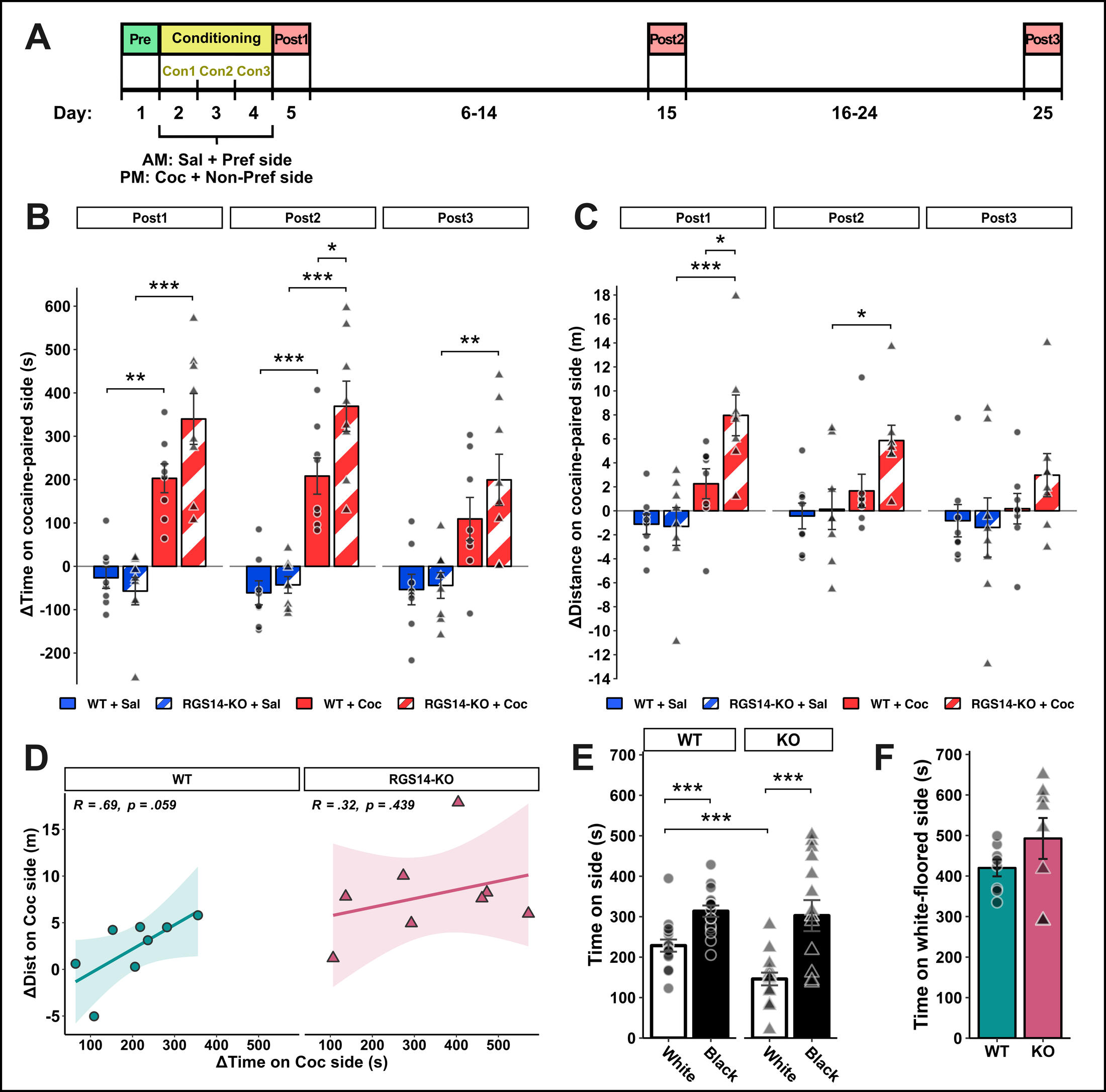
Endogenous RGS14 blunts cocaine-induced emotionally motivated behaviors in female mice
Bramlett, S. N., Foster, S. L., Weinshenker, D., & Hepler, J. R.
Addiction Neuroscience, 15, 100203
2024 First Author
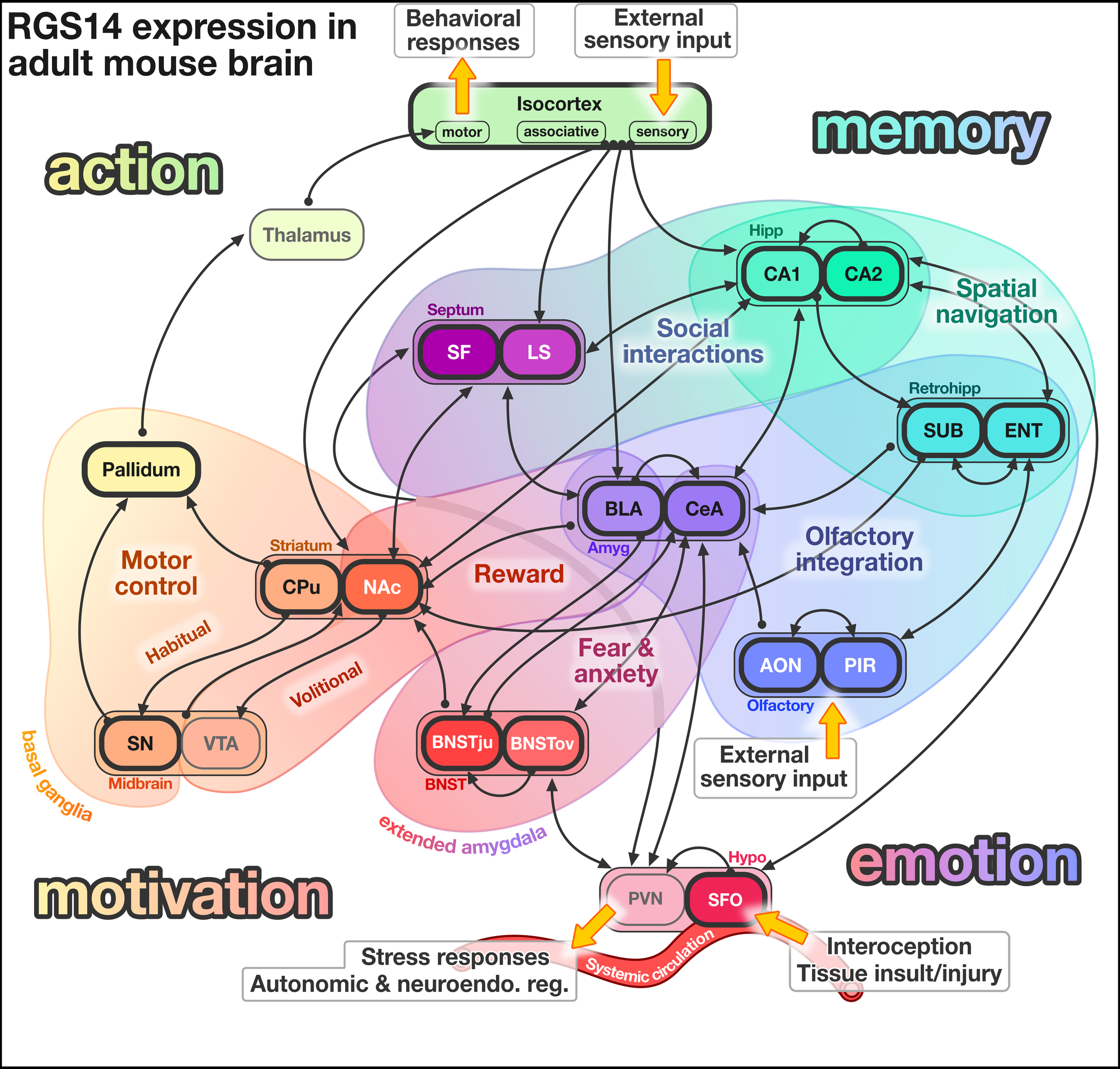
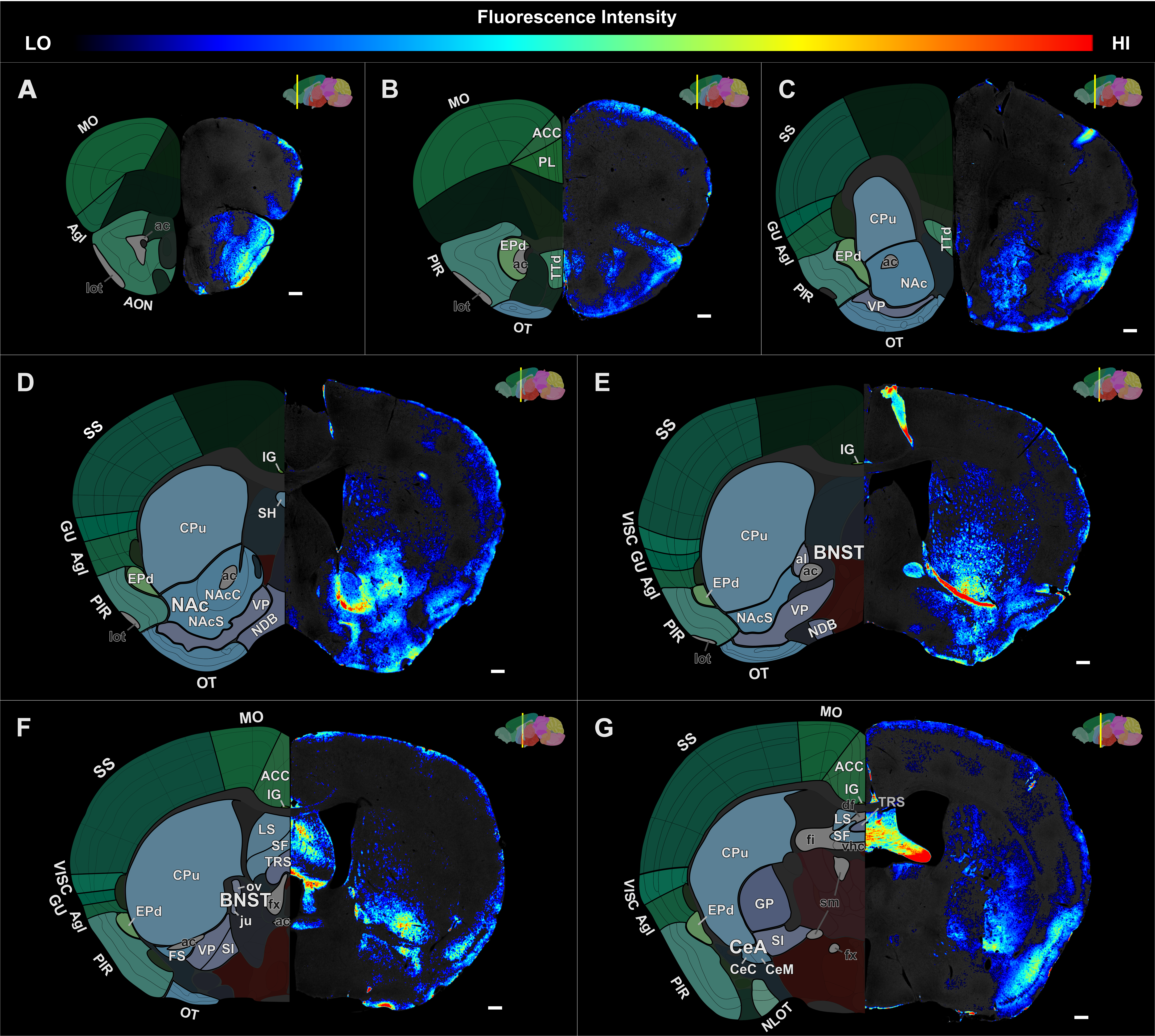
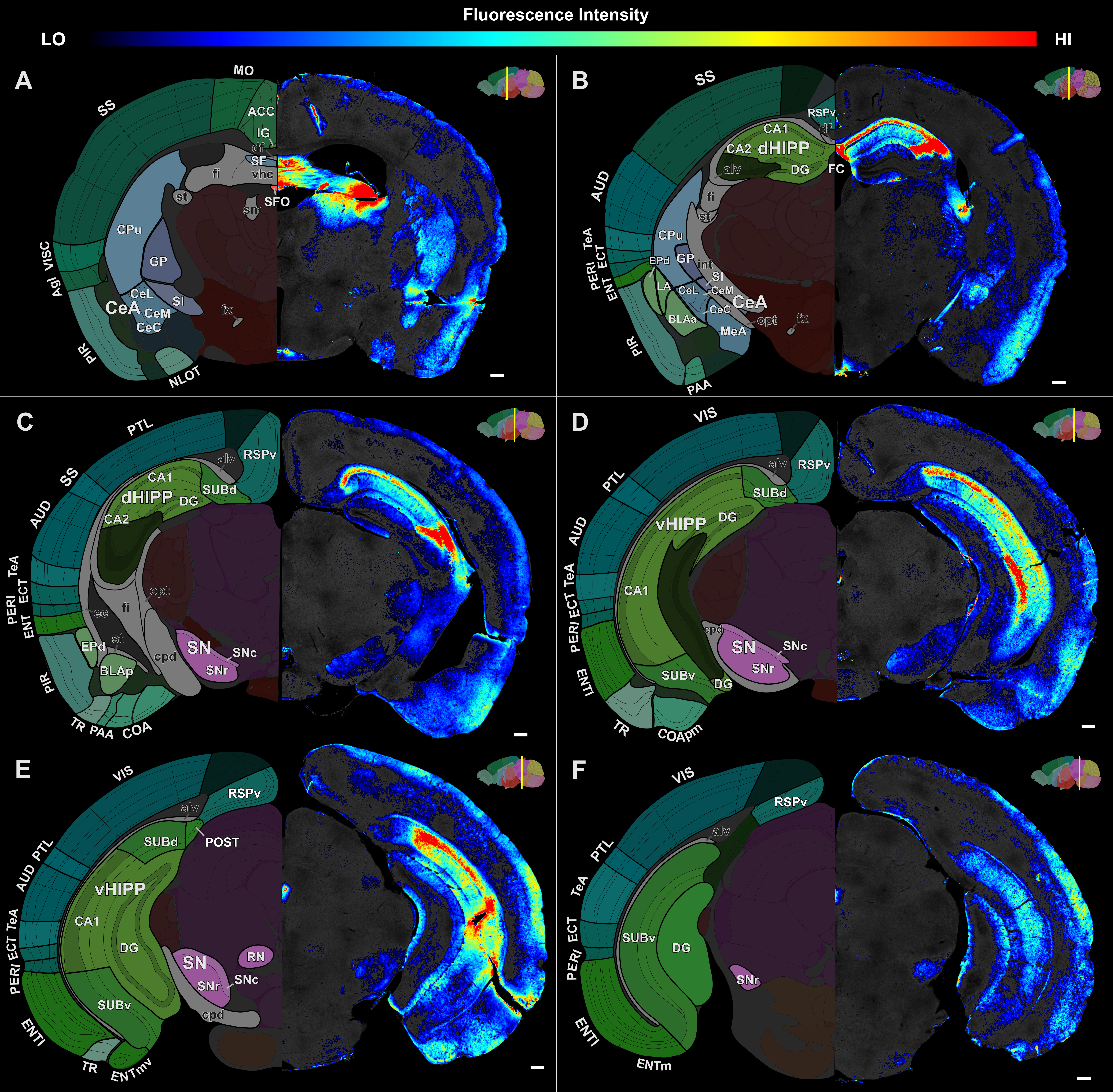
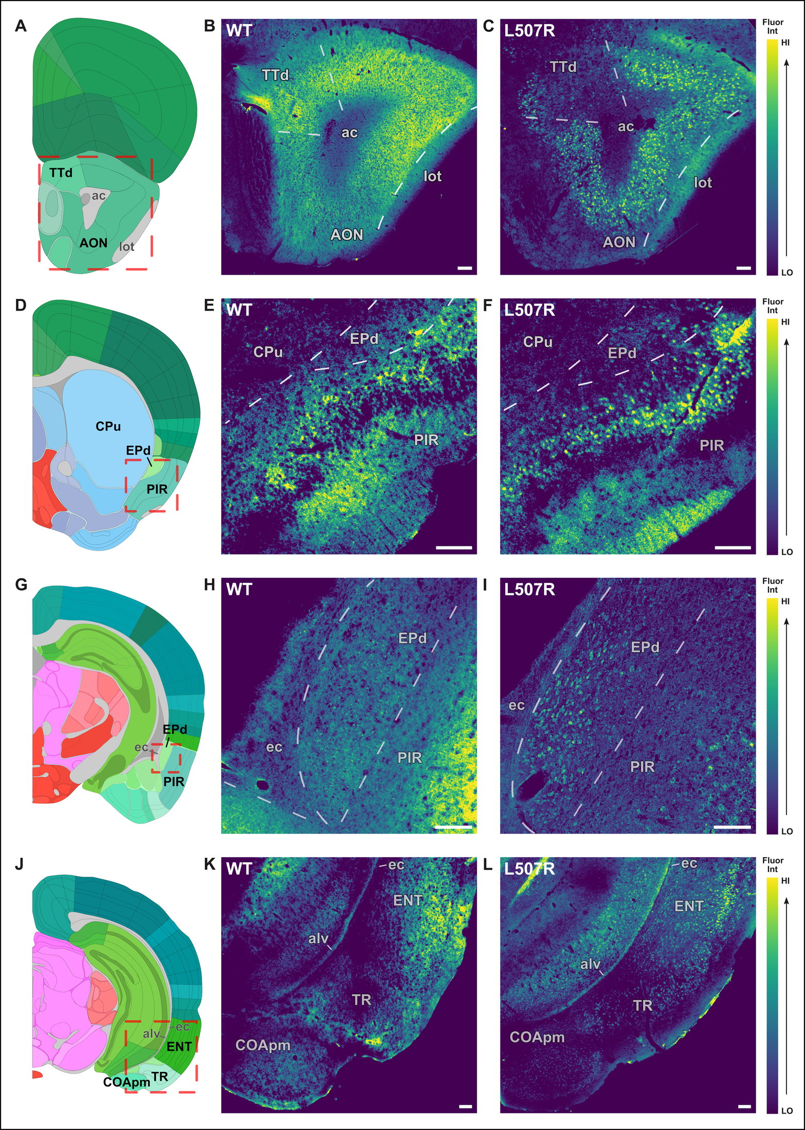
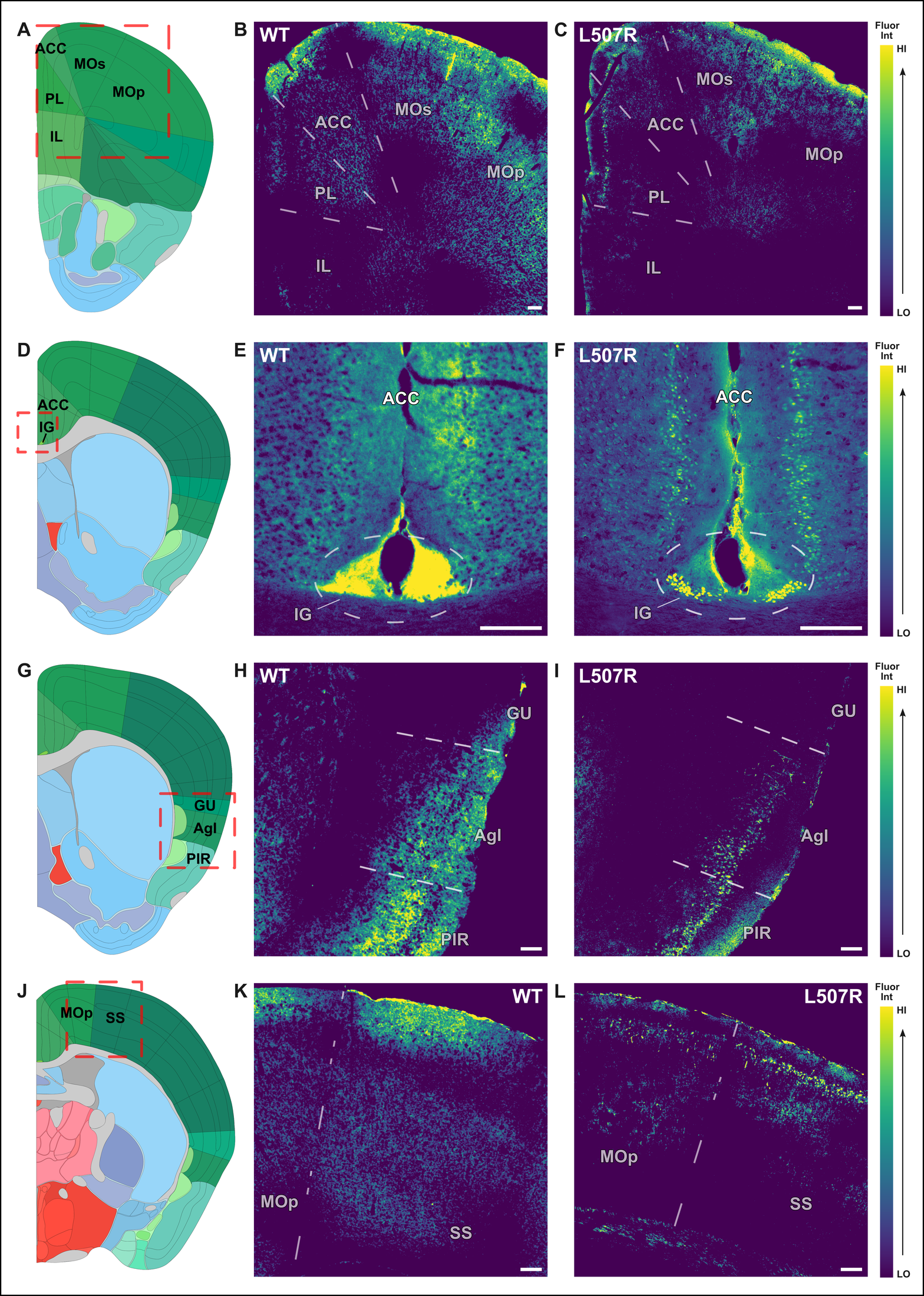
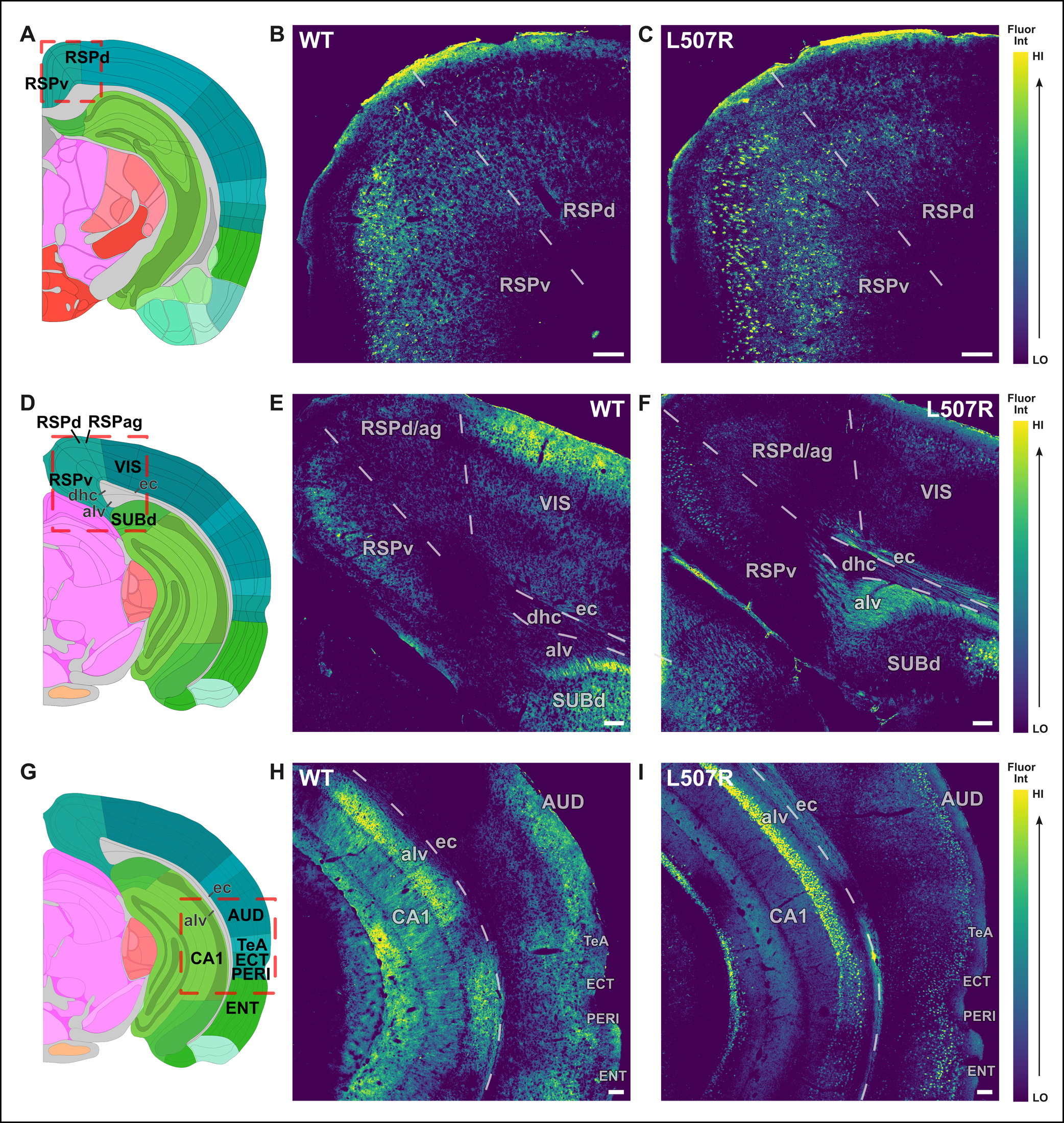
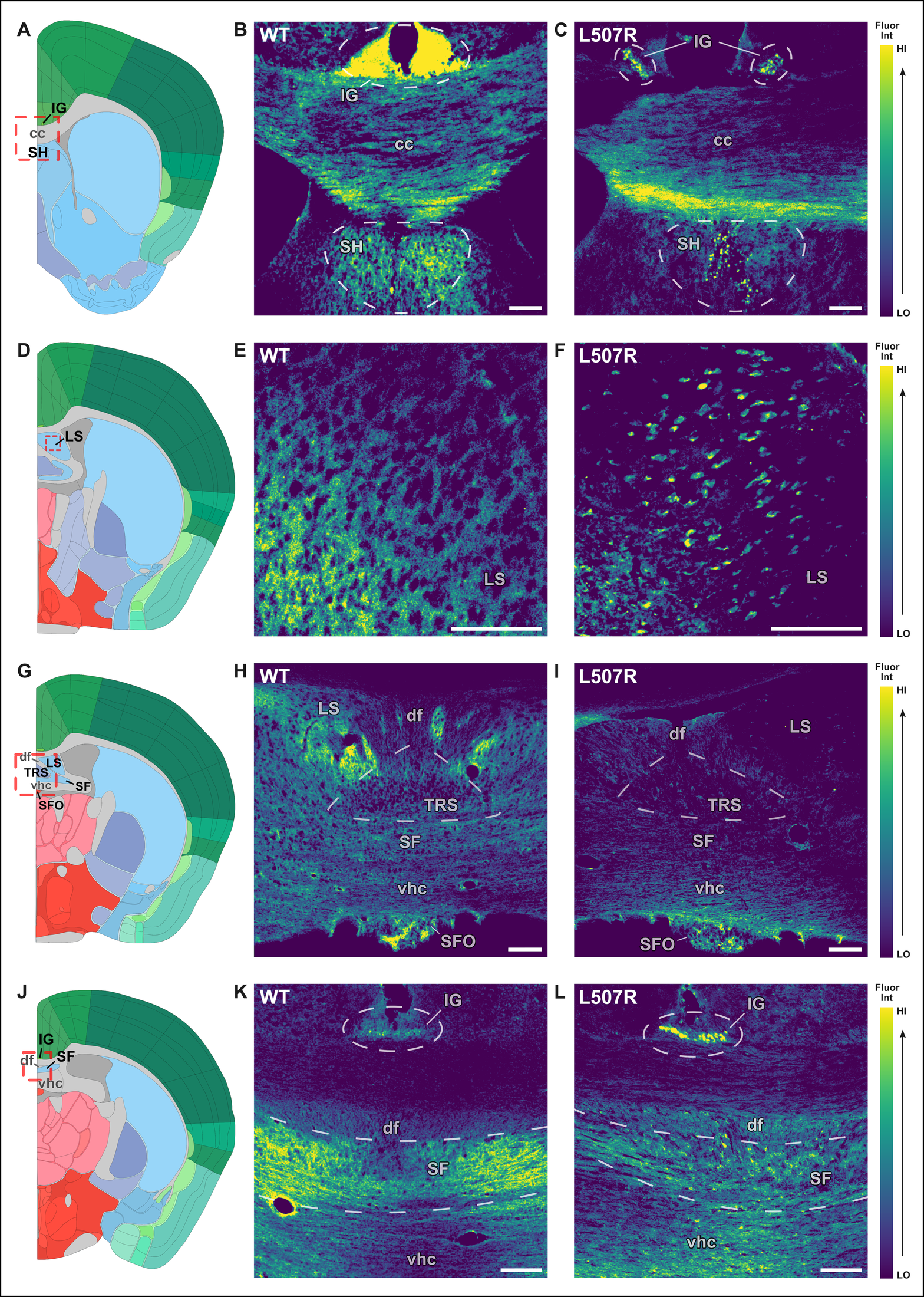
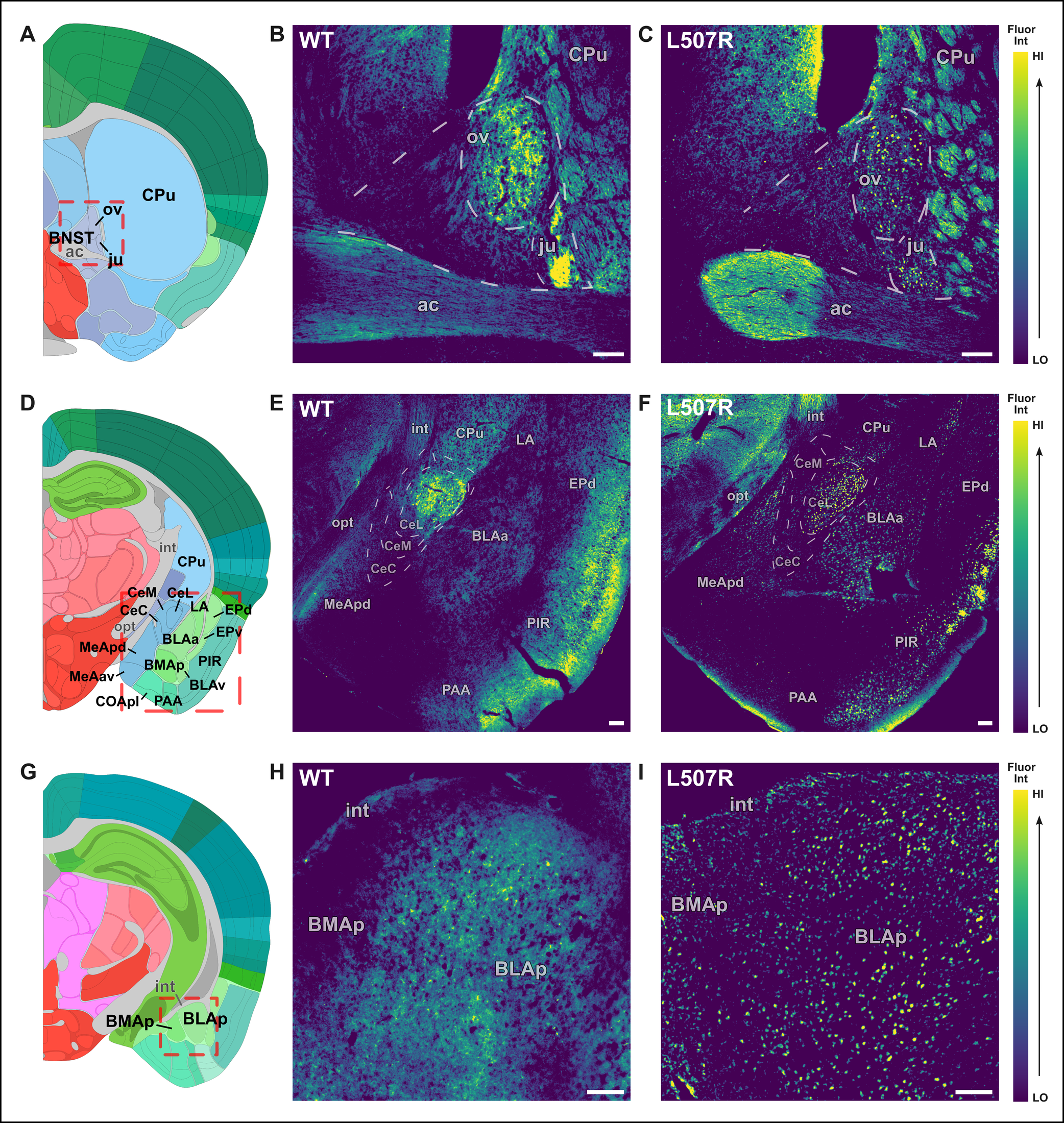
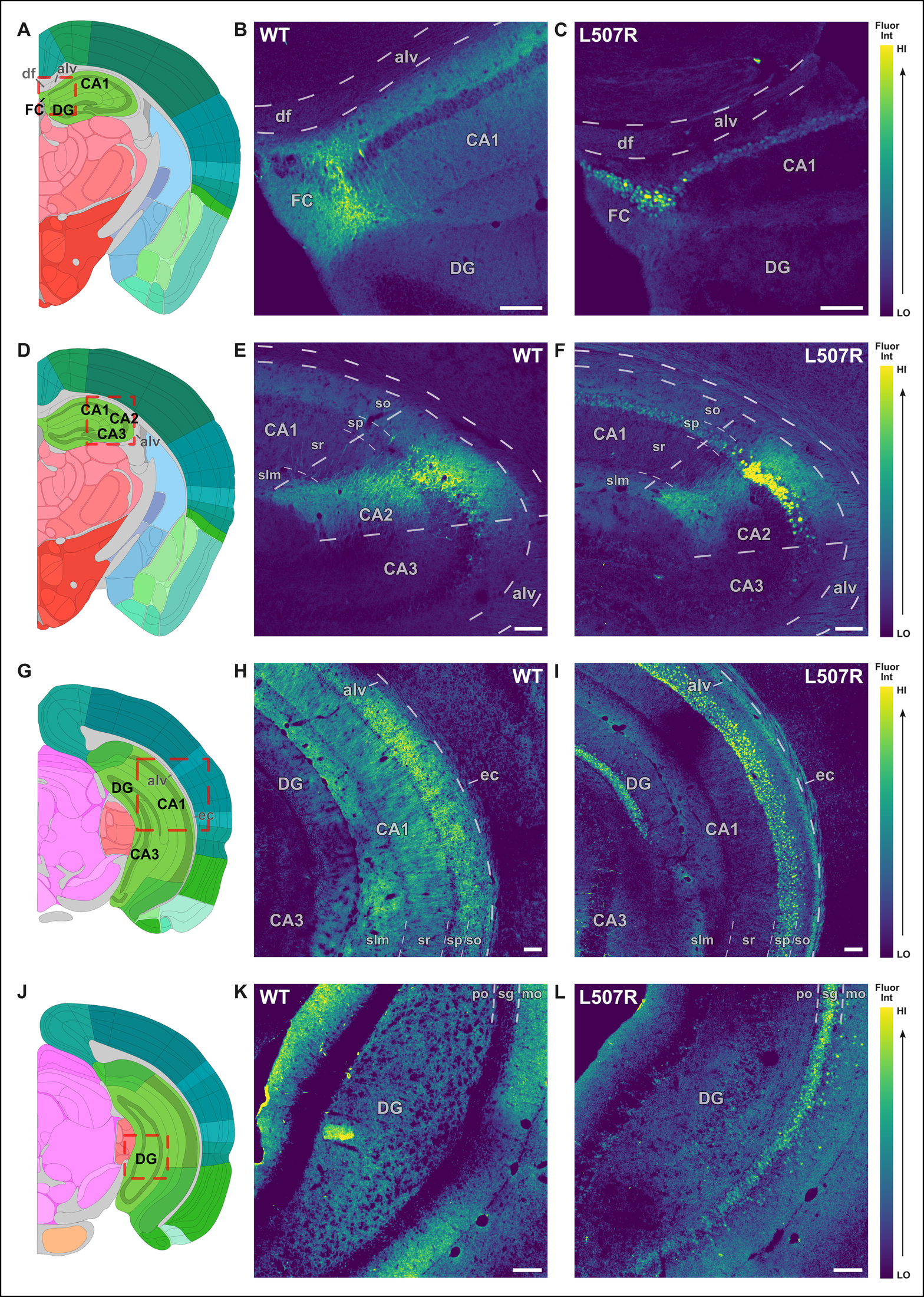
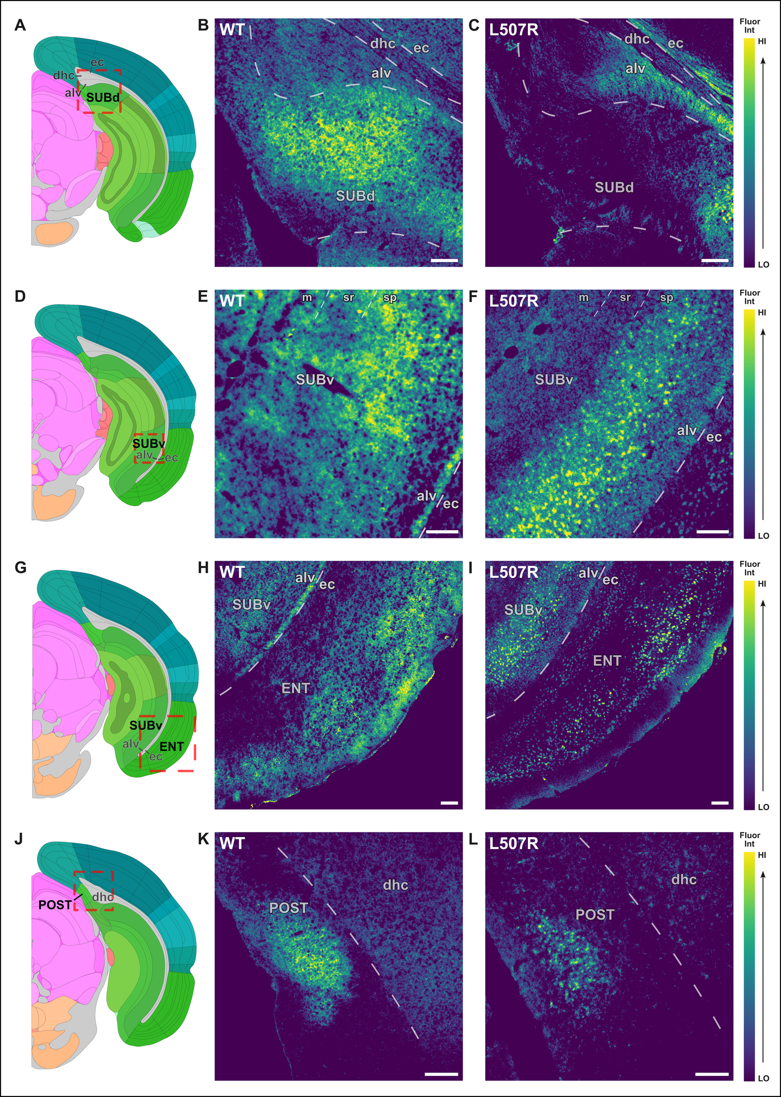
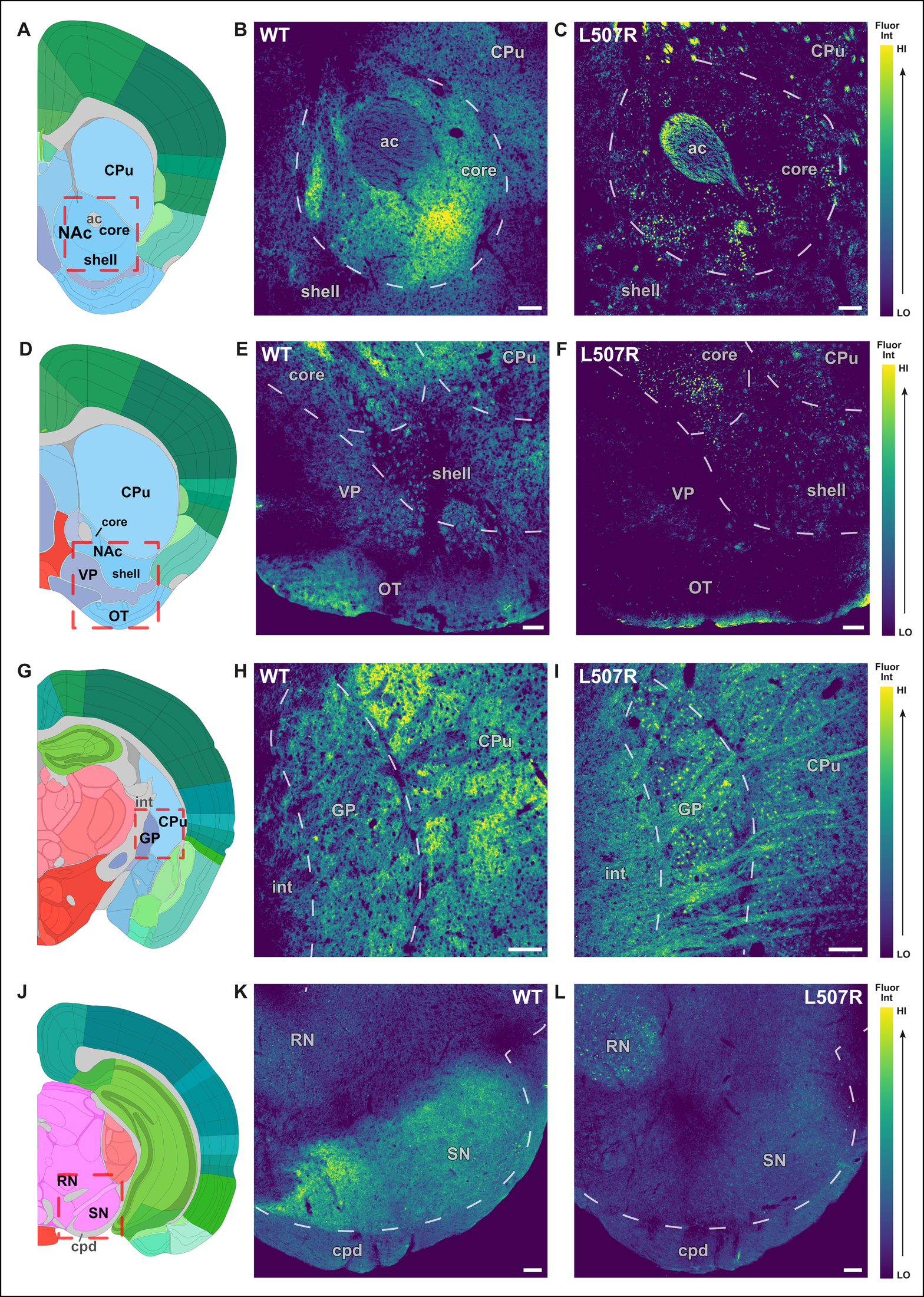
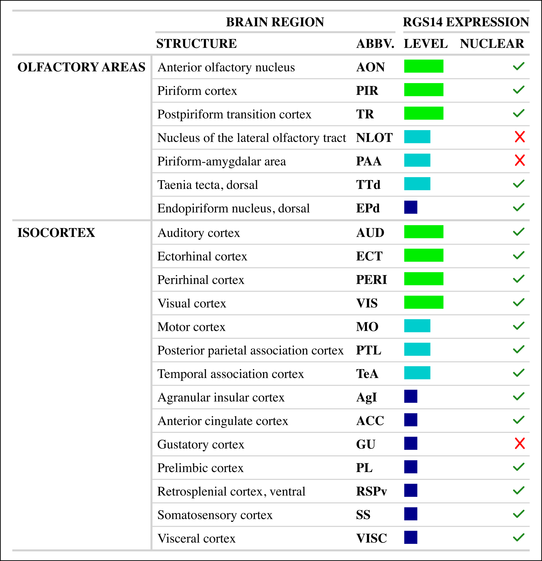
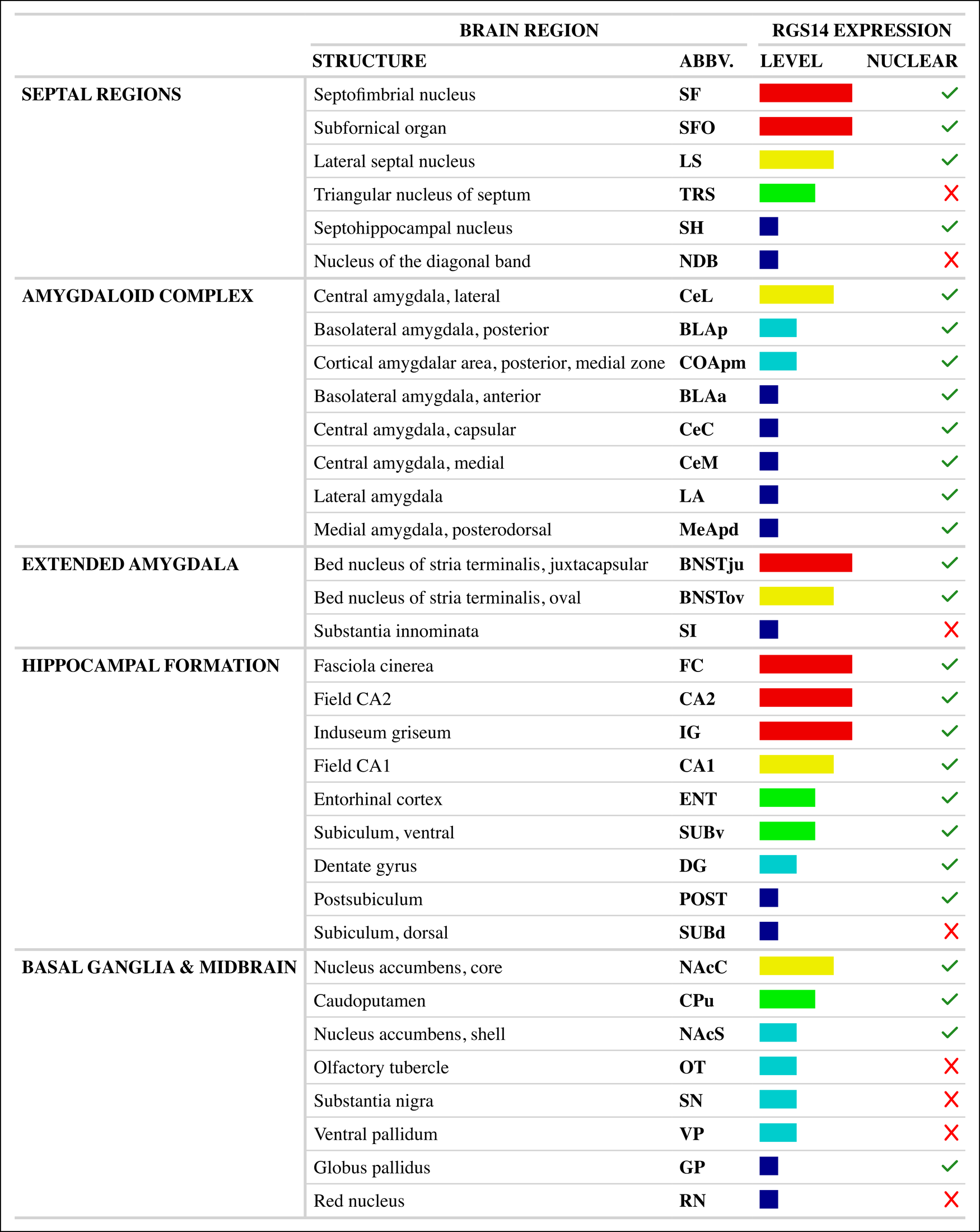
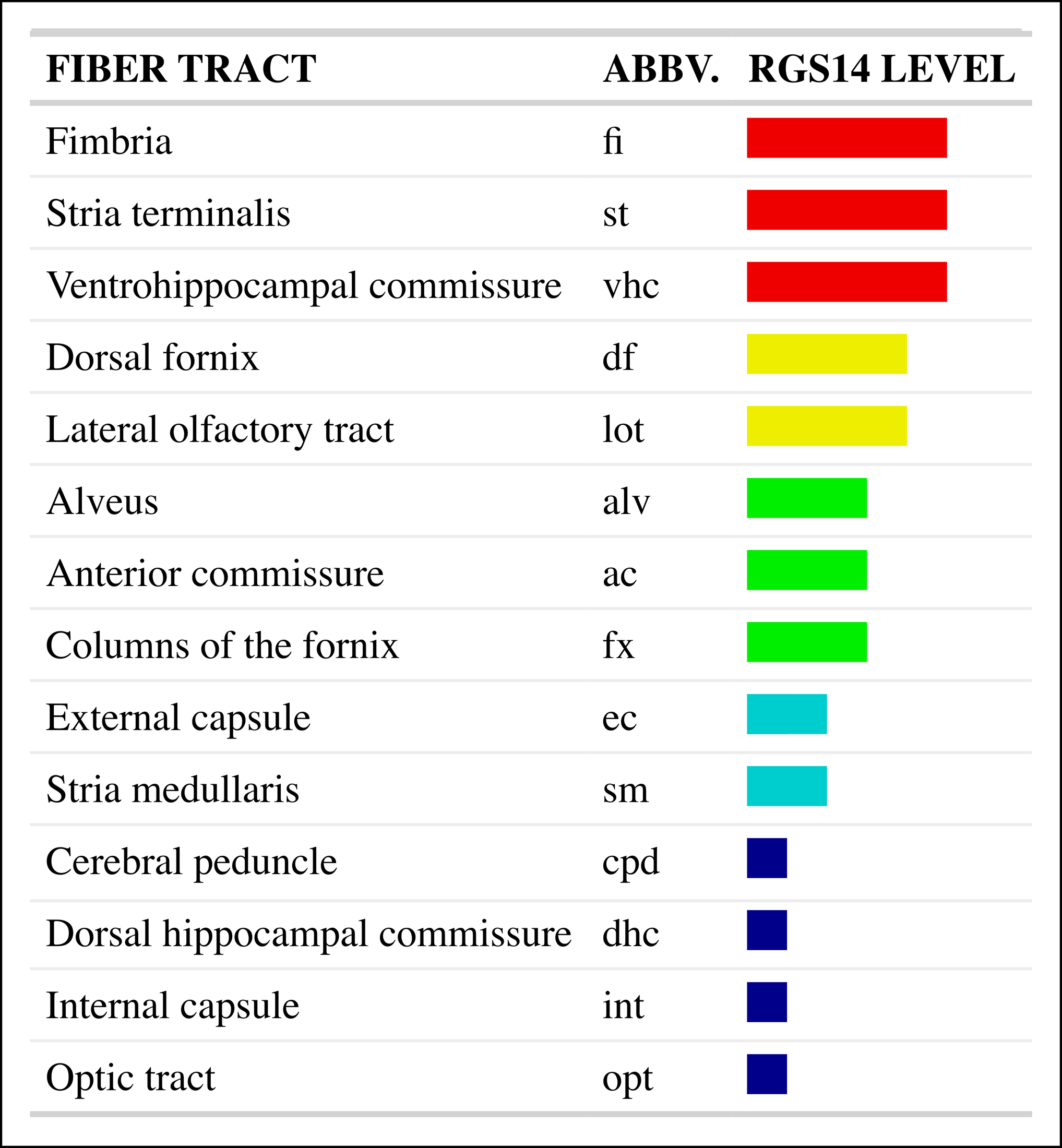
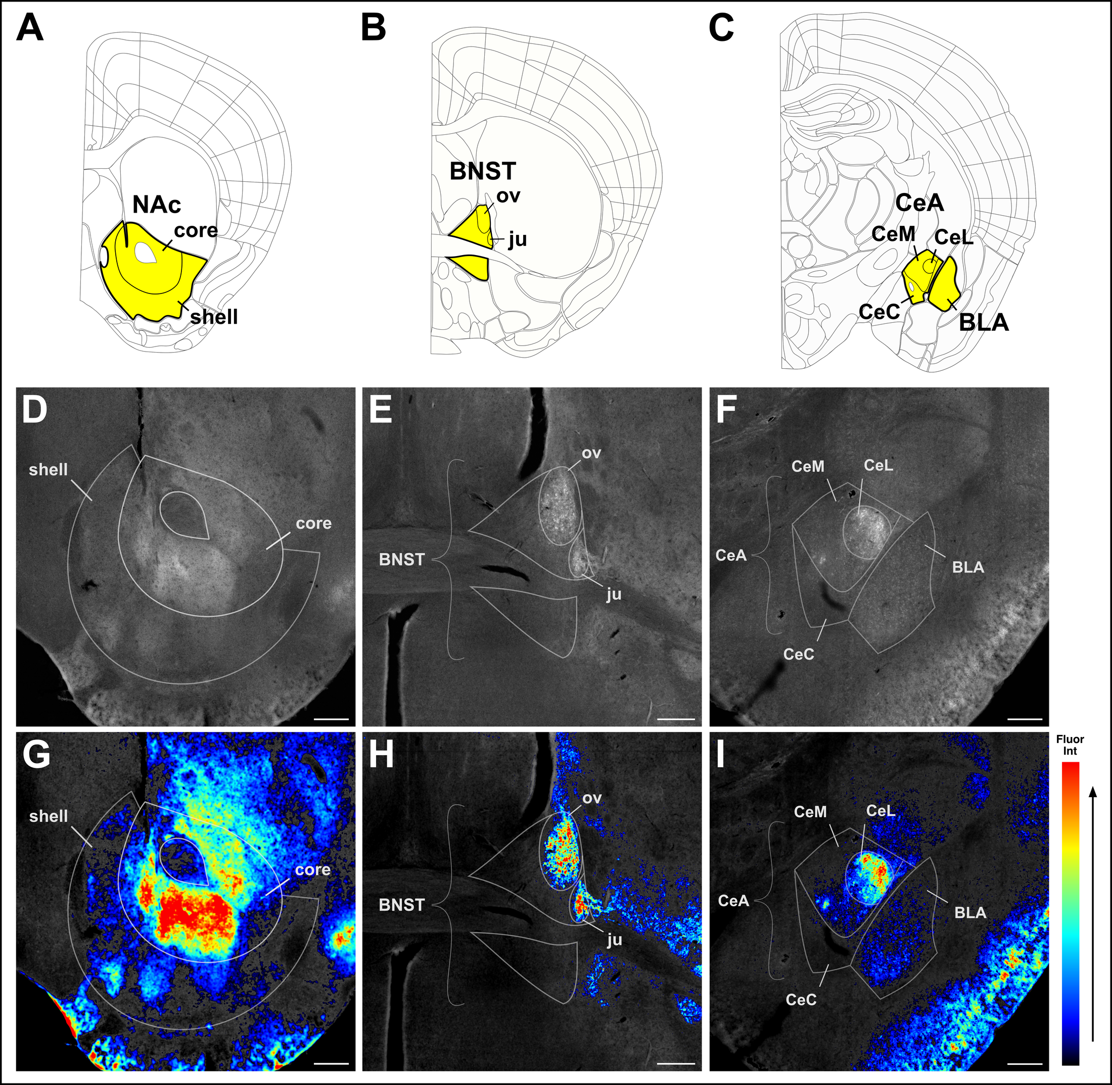
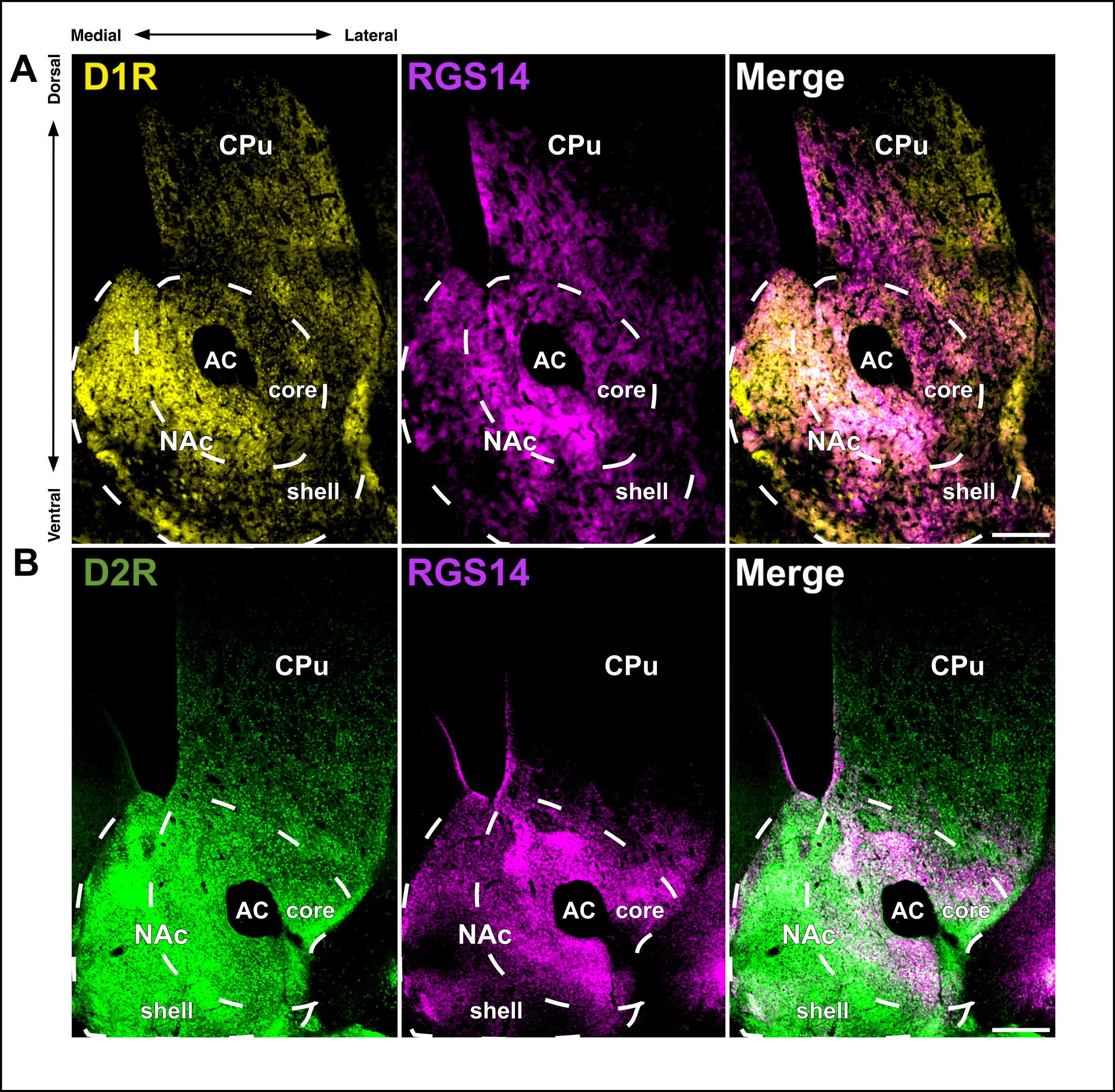
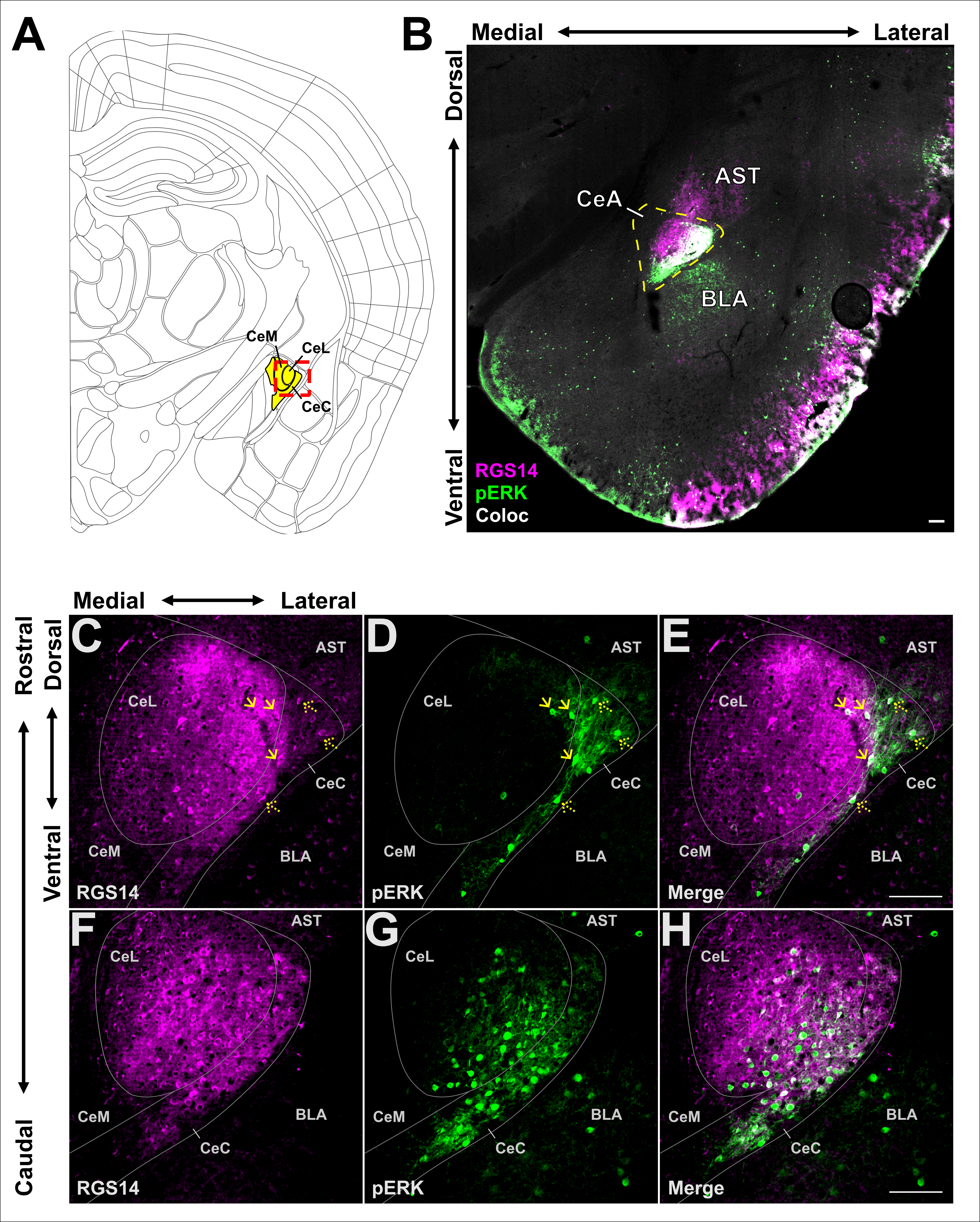
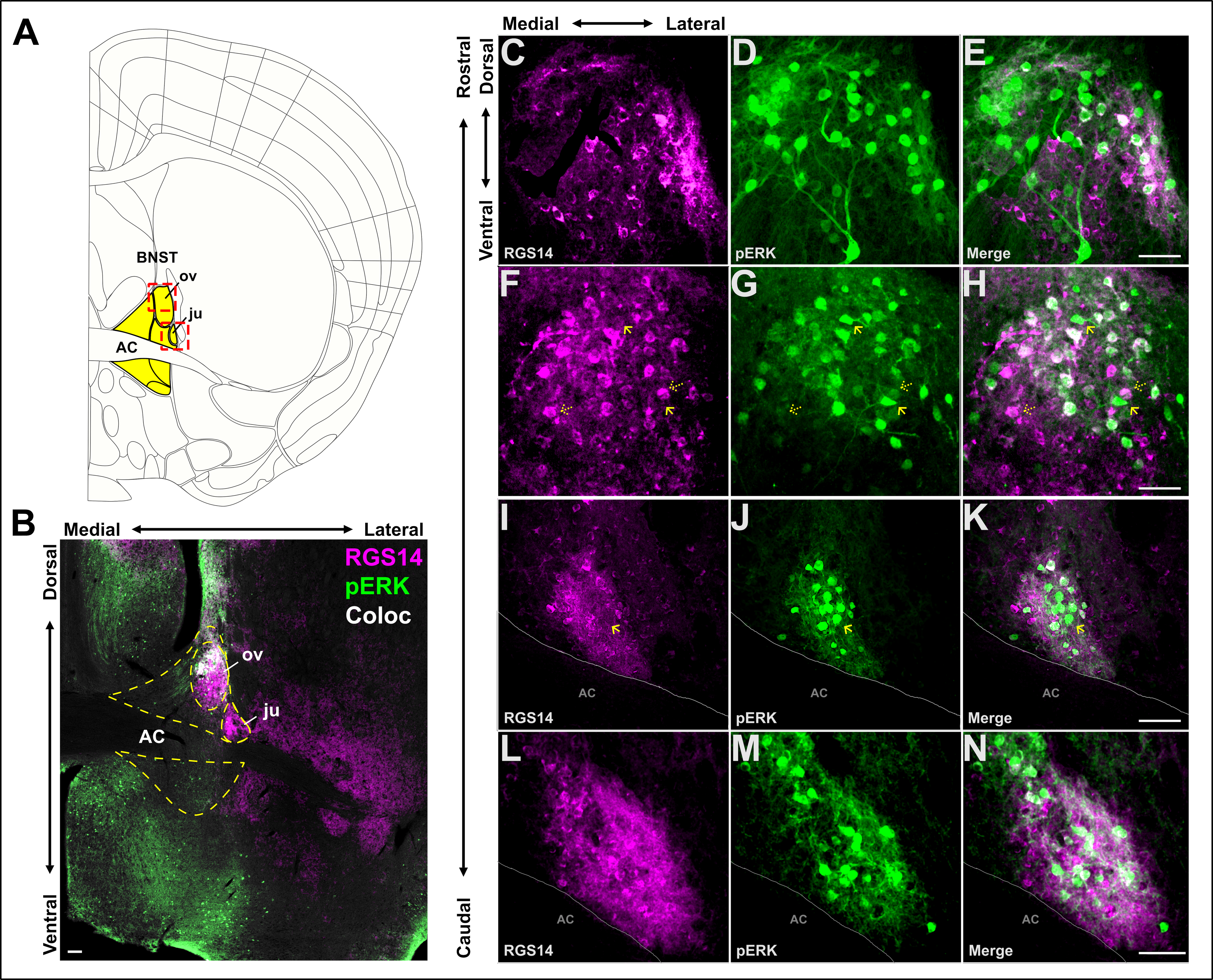
RGS14 protein expression profile in the adult mouse brain
Bramlett, S. N., Fitzmaurice, S. M., Harbin, N. H., Yan, W., Bandlamudi, C., Van Doorn, G. E., Smith, Y., & Hepler, J. R.
European Journal of Neuroscience · DOI: 10.1111/ejn.16592
2023
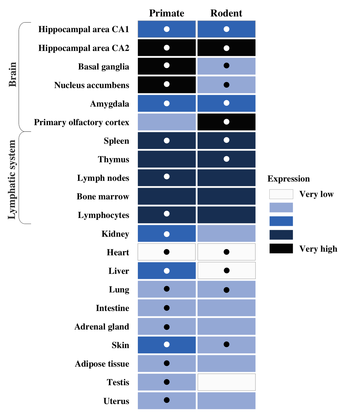
RGS14 expression in CA2 hippocampus, amygdala, and basal ganglia: implications for human brain physiology and disease
Montanez-Miranda, C., Bramlett, S. N., & Hepler, J. R.
Hippocampus · DOI: 10.1002/hipo.23492
2021
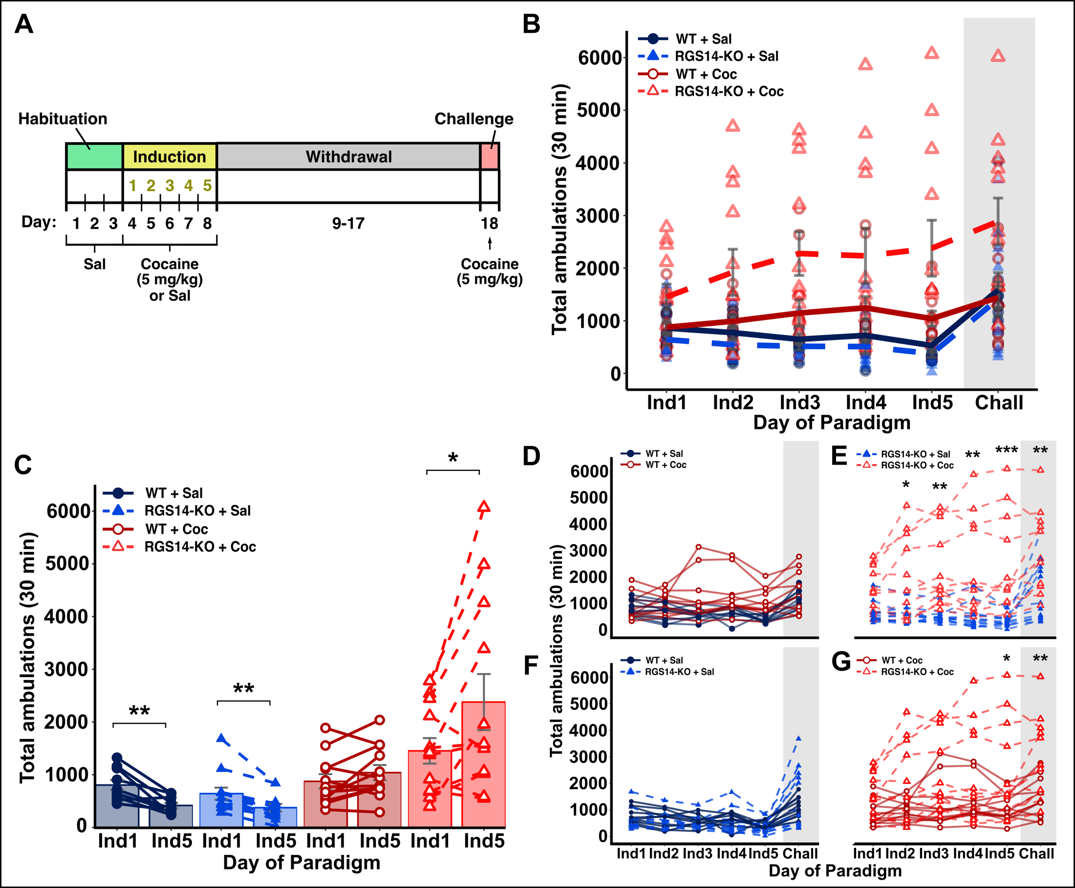
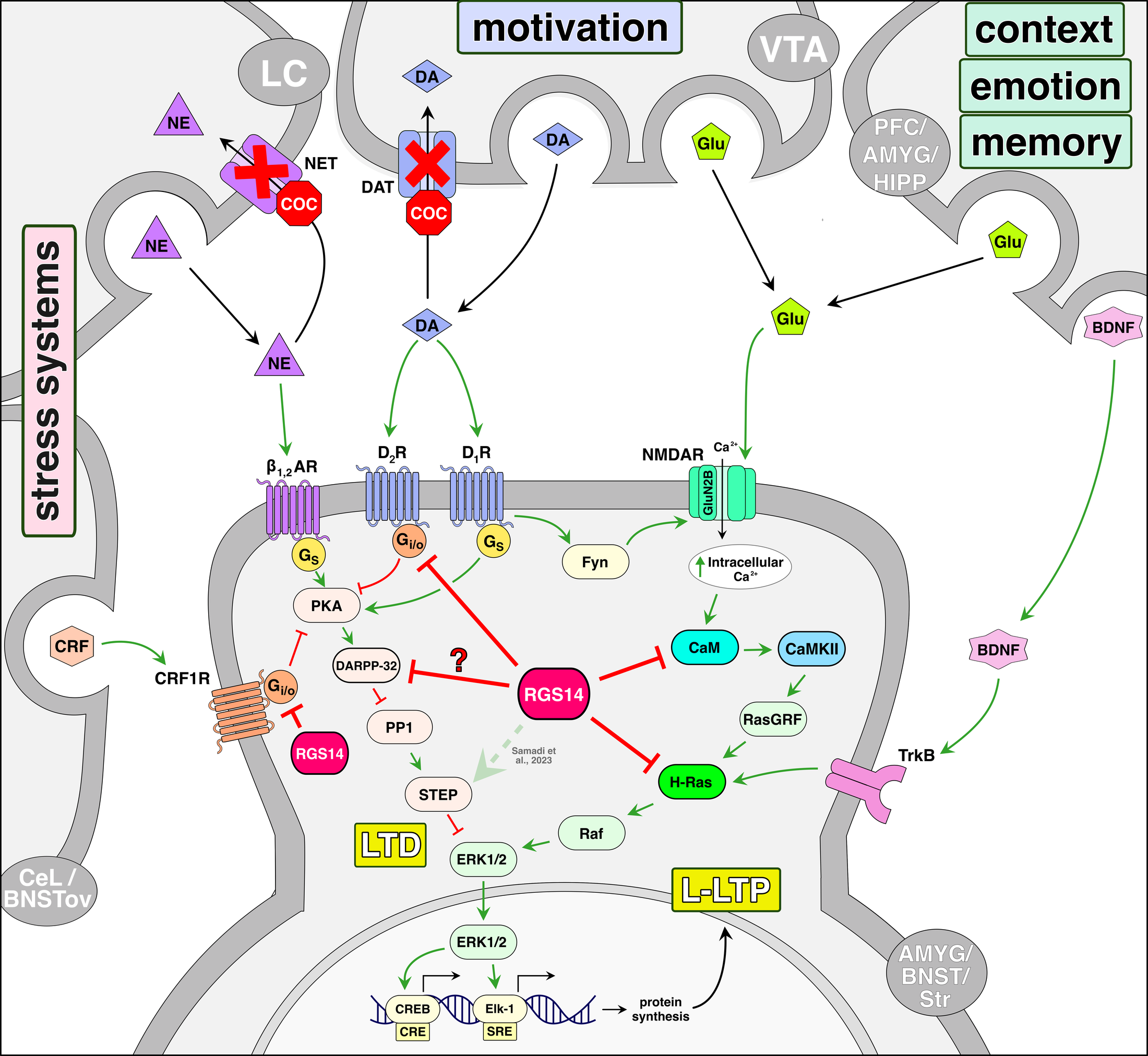
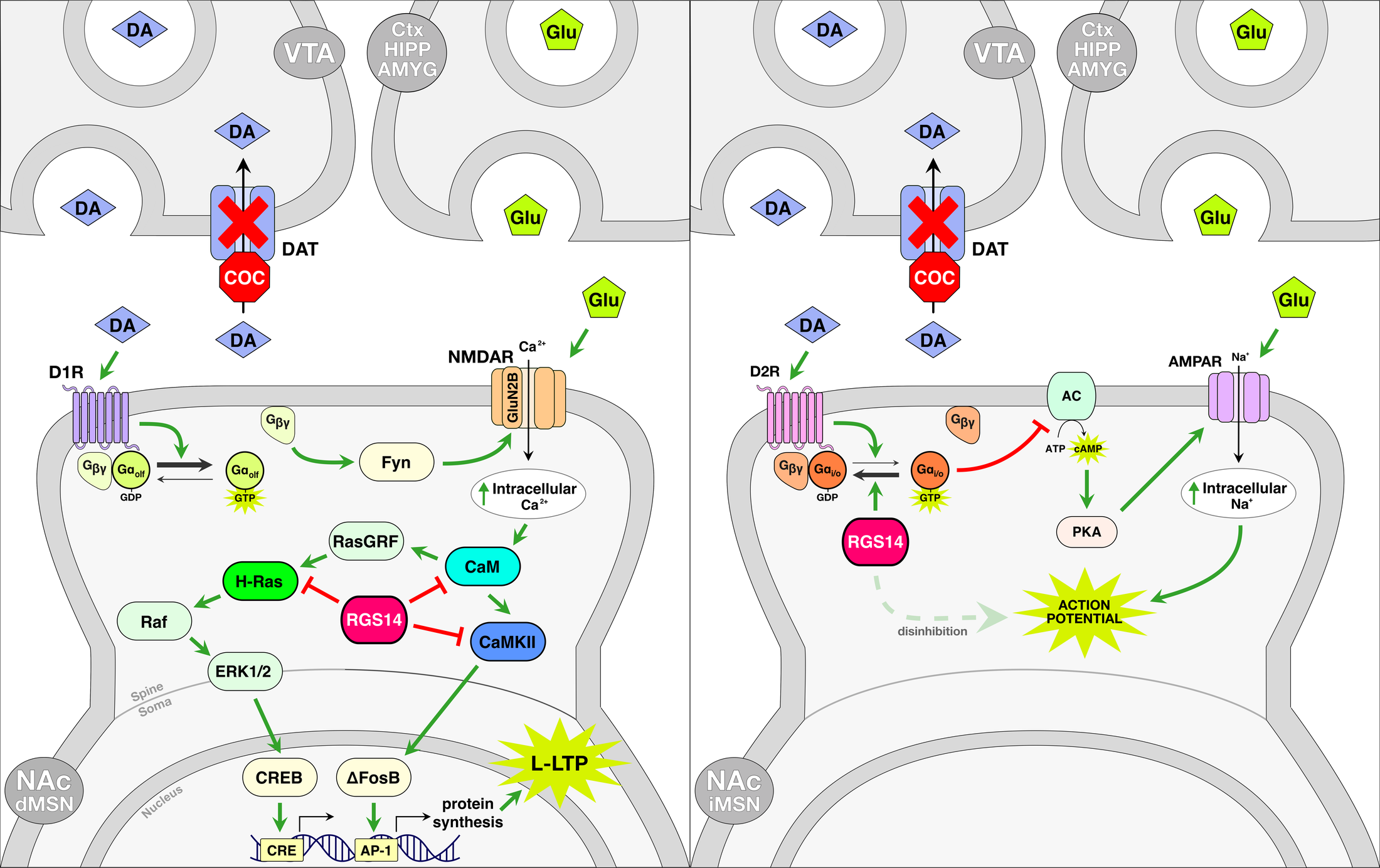
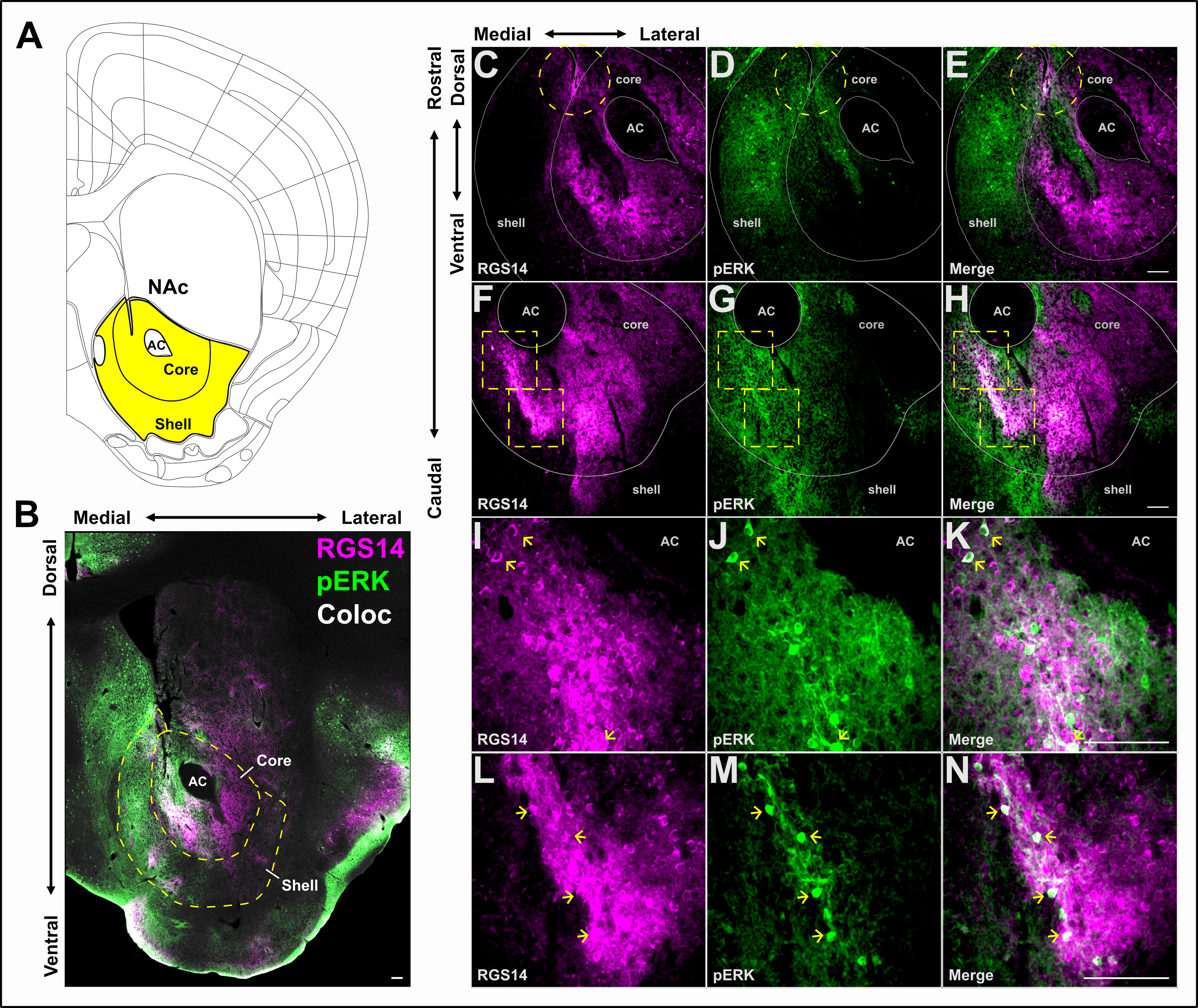
RGS14 modulates locomotor behavior and ERK signaling induced by environmental novelty and cocaine within discrete limbic structures
Foster, S. L., Lustberg, D. J., Harbin, N. H., Bramlett, S. N., Hepler, J. R., & Weinshenker, D.
Psychopharmacology, 238(10), 2755–2773
2021
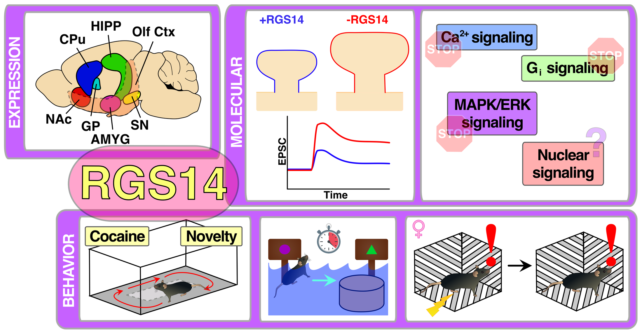
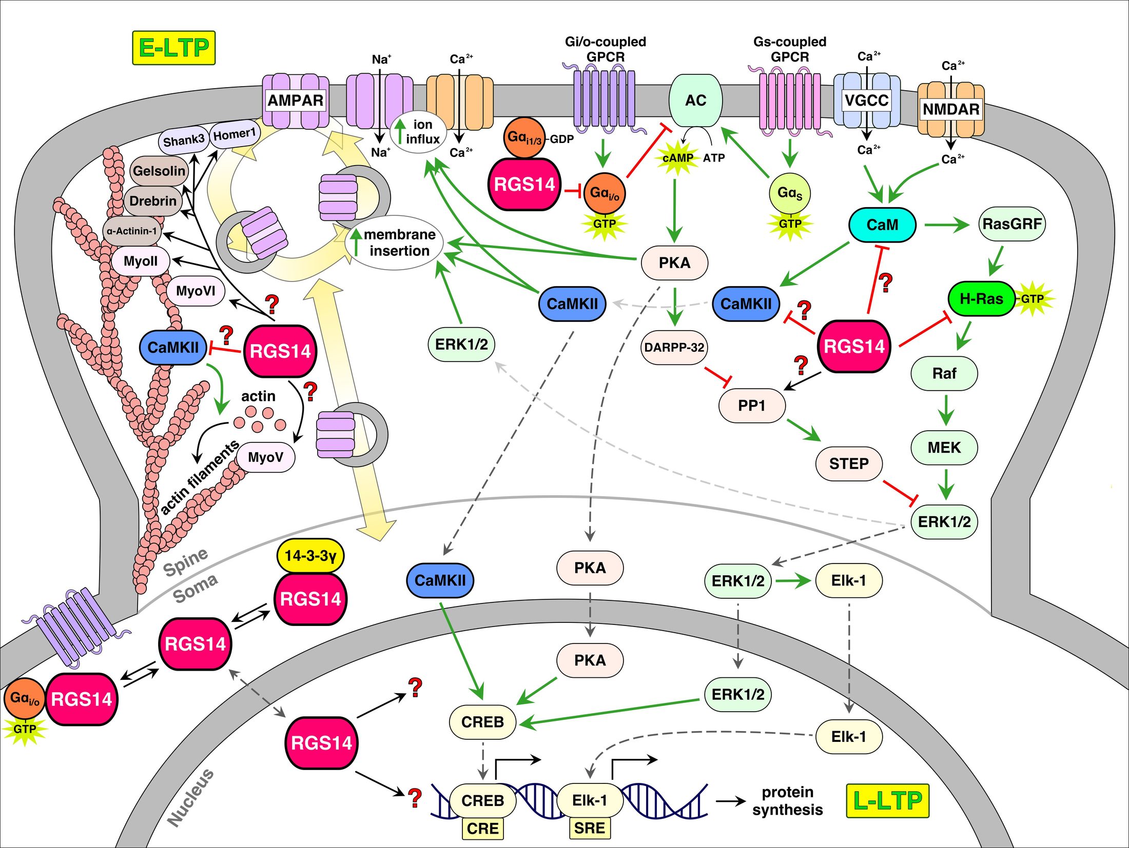
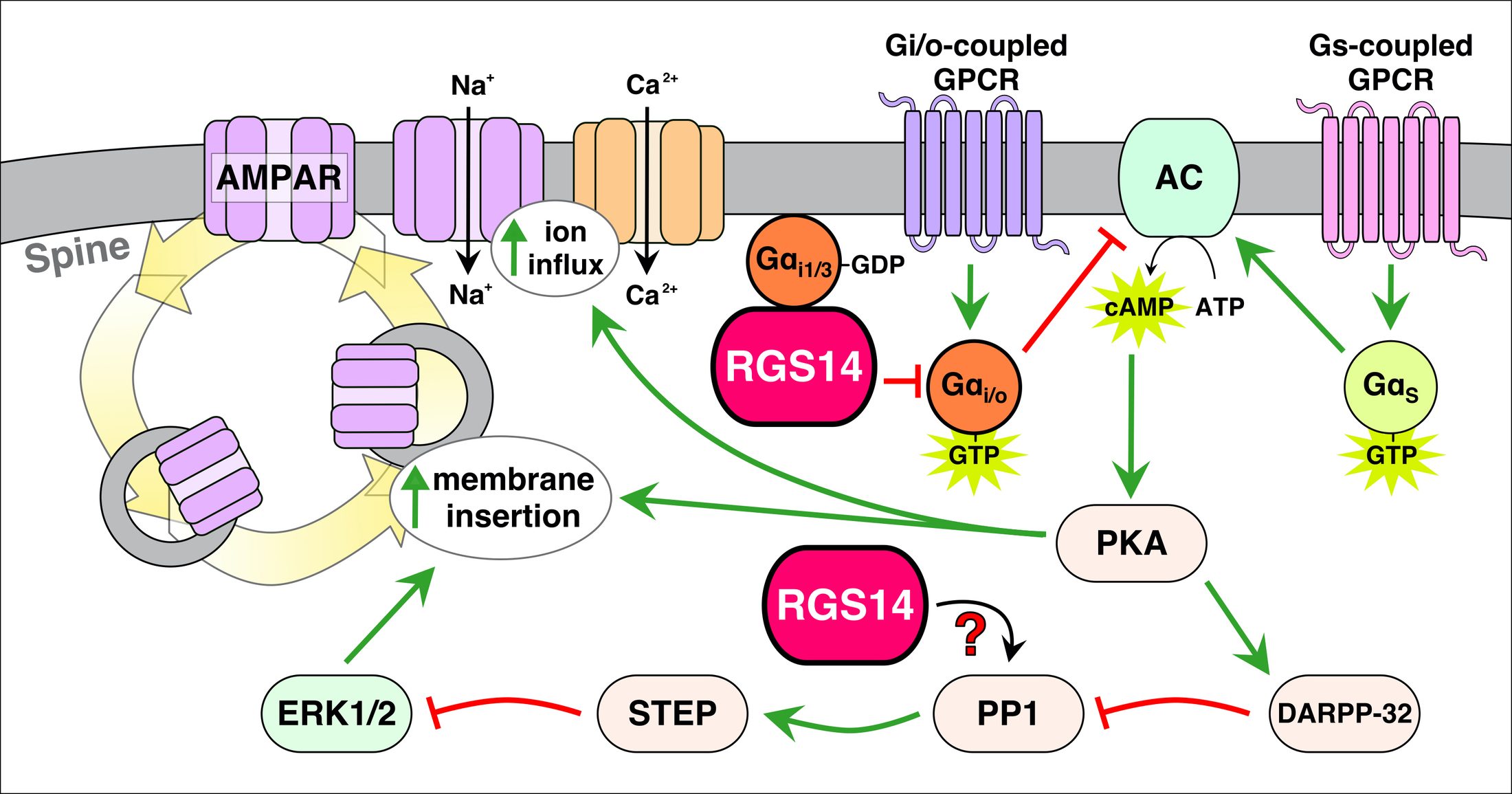
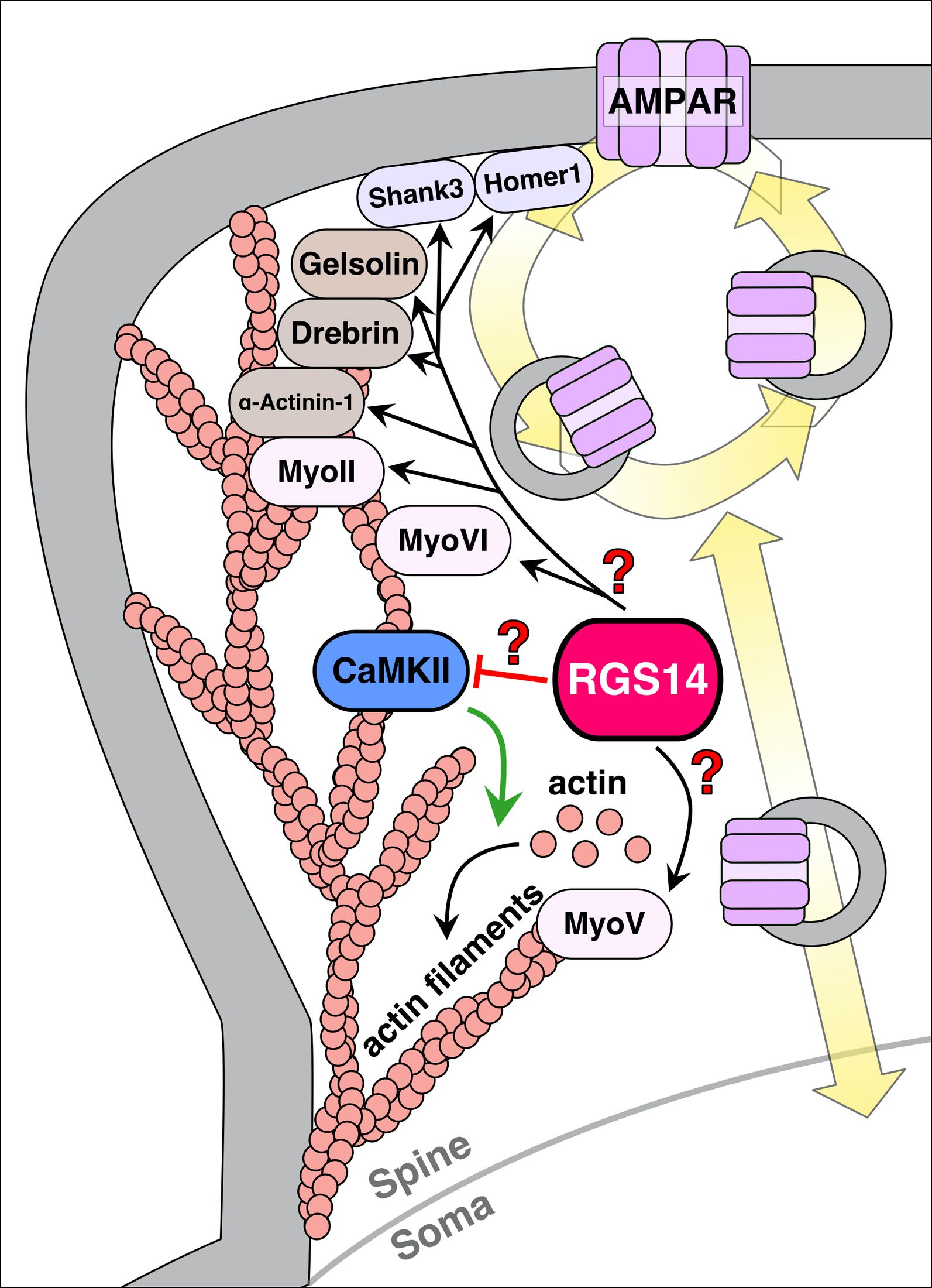
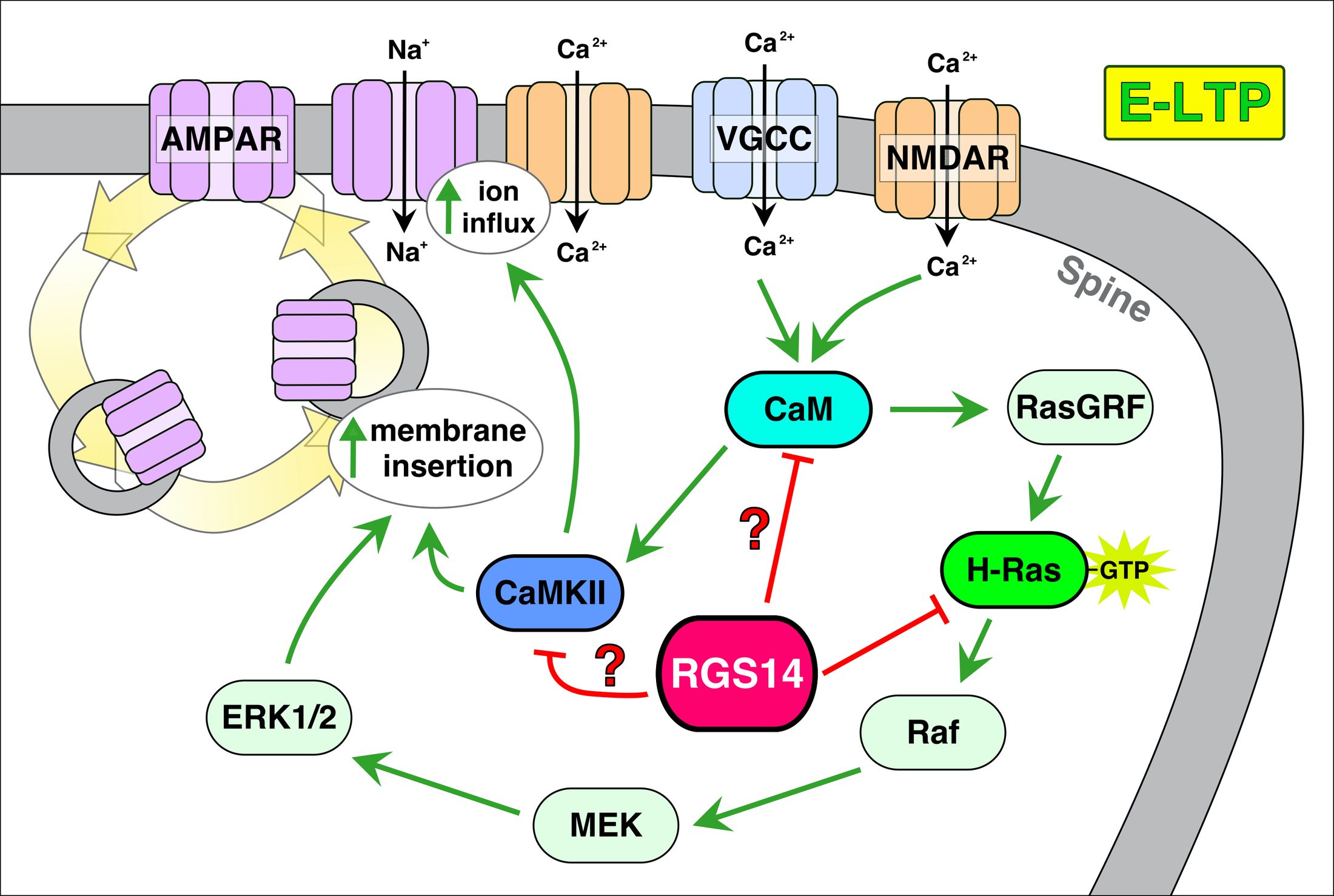
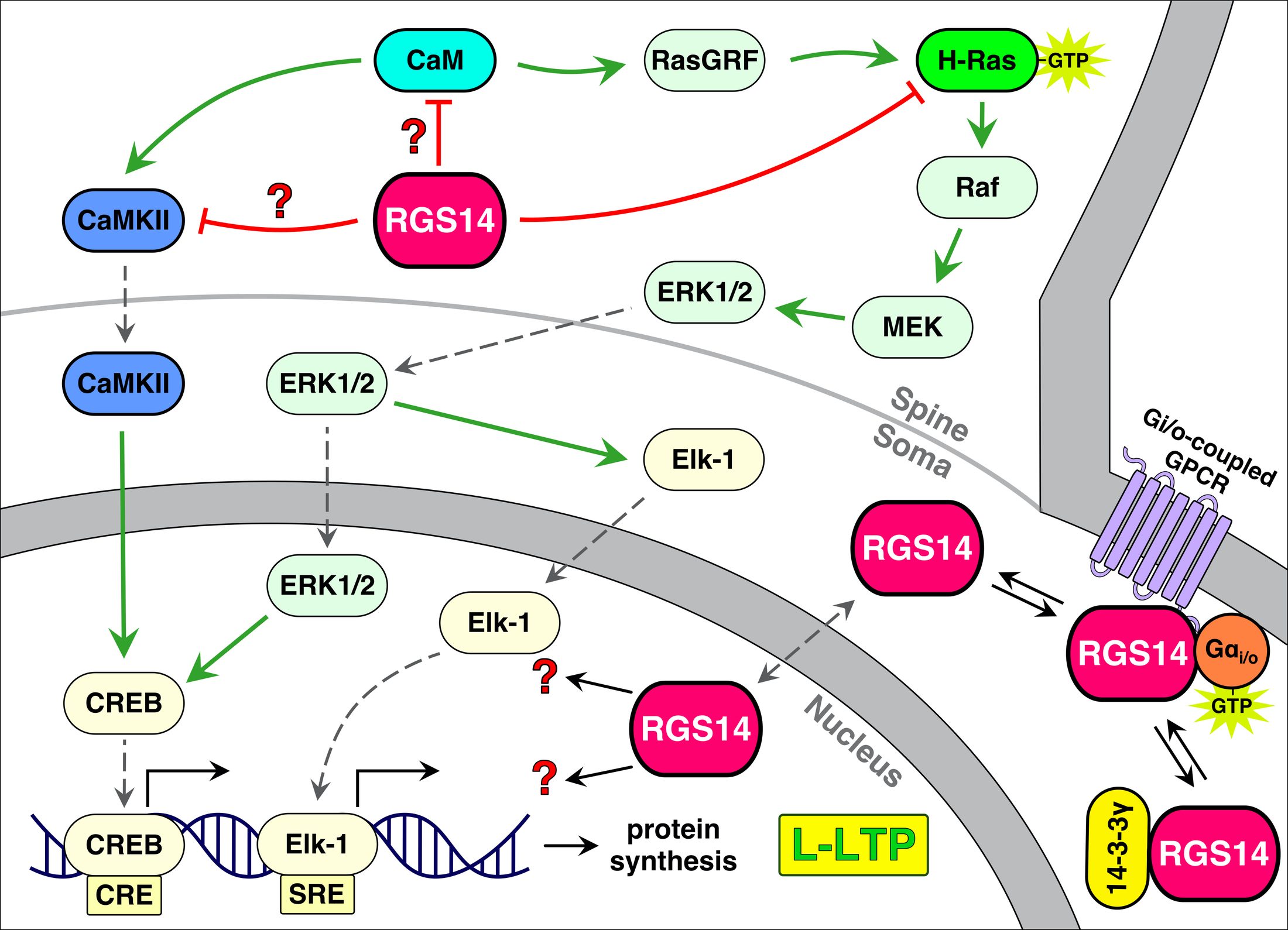
RGS14 regulation of post-synaptic signaling and spine plasticity in brain
Harbin, N. H., Bramlett, S. N., Montanez-Miranda, C., Terzioglu, G., & Hepler, J. R.
International Journal of Molecular Sciences, 22(13), 6823
2019
Effects of early life stress on cocaine self-administration in post-pubertal male and female rhesus macaques
Wakeford, A. G. P., Morin, E. L., Bramlett, S. N., Howell, B. R., McCormack, K. M., Meyer, J. S., Nader, M. A., Sanchez, M. M., & Howell, L. L.
Psychopharmacology, 236, 2785–2796
2018
A review of nonhuman primate models of early life stress and adolescent drug abuse
Wakeford, A. G. P., Morin, E. L., Bramlett, S. N., Howell, L. L., & Sanchez, M. M.
Neurobiology of Stress, 9, 188–198
Contact

Let's Work Together

Available for freelance scientific illustration and science communication. Also happy to connect about research or just say hello. Reach me at SciOpsDesign@gmail.com or sara.n.bramlett@gmail.com.